From the Department of Radiology, Juntendo University.
MR Applications and Workflow, GE Healthcare Japan, Tokyo, Japan.
Invest Radiol. 2021 May 1;56(5):292-300. doi: 10.1097/RLI.0000000000000744.
The aims of this study were to develop an accelerated multiparametric magnetic resonance imaging method based on 3D-quantification using an interleaved Look-Locker acquisition sequence with a T2 preparation pulse (3D-QALAS) combined with compressed sensing (CS) and to evaluate the effect of CS on the quantitative mapping, tissue segmentation, and quality of synthetic images.
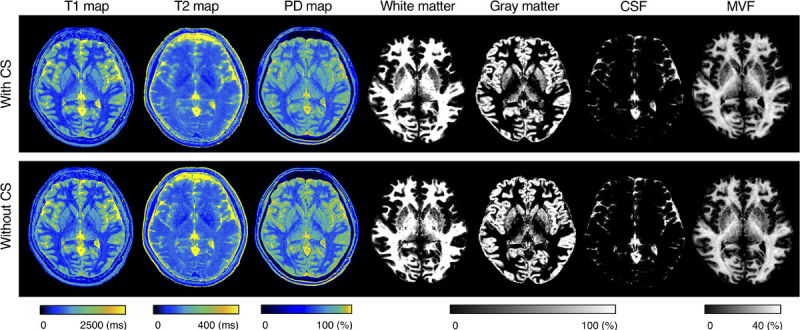
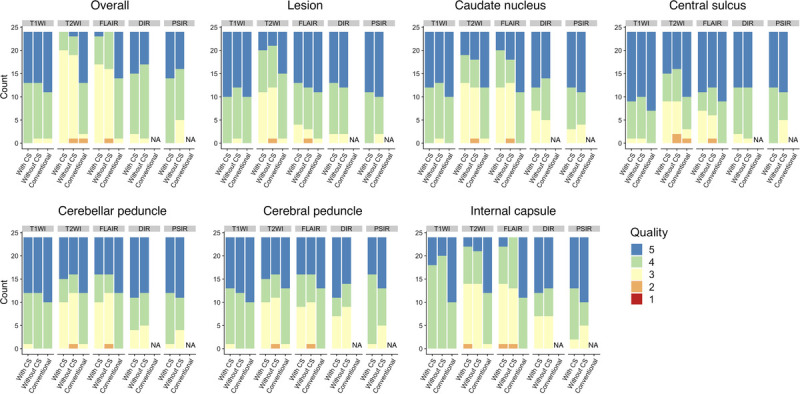
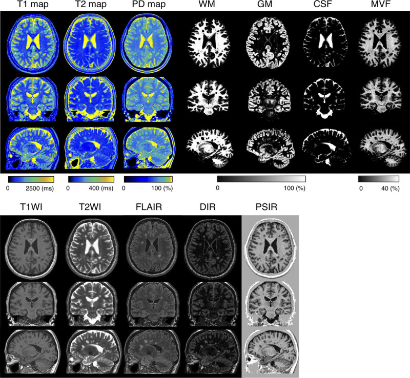
A magnetic resonance imaging system phantom, containing multiple compartments with standardized T1, T2, and proton density (PD) values; 10 healthy volunteers; and 12 patients with multiple sclerosis were scanned using the 3D-QALAS sequence with and without CS and conventional contrast-weighted imaging. The scan times of 3D-QALAS with and without CS were 5:56 and 11:11, respectively. For healthy volunteers, brain volumetry and myelin estimation were performed based on the measured T1, T2, and PD. For patients with multiple sclerosis, the mean T1, T2, PD, and the amount of myelin in plaques and contralateral normal-appearing white matter (NAWM) were measured. Simple linear regression analysis and Bland-Altman analysis were performed for each metric obtained from the datasets with and without CS. To compare overall image quality and structural delineations on synthetic and conventional contrast-weighted images, case-control randomized reading sessions were performed by 2 neuroradiologists in a blinded manner.
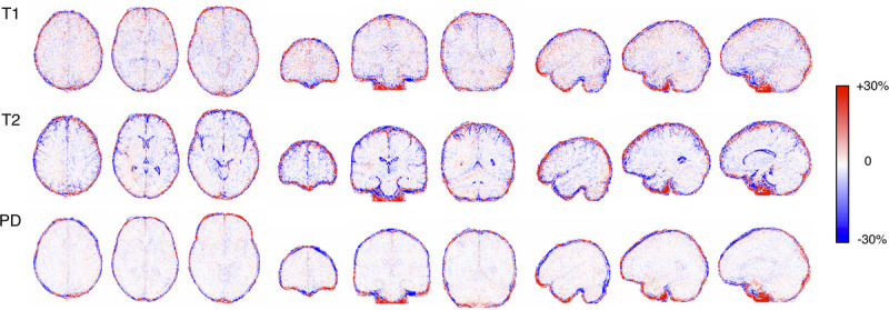
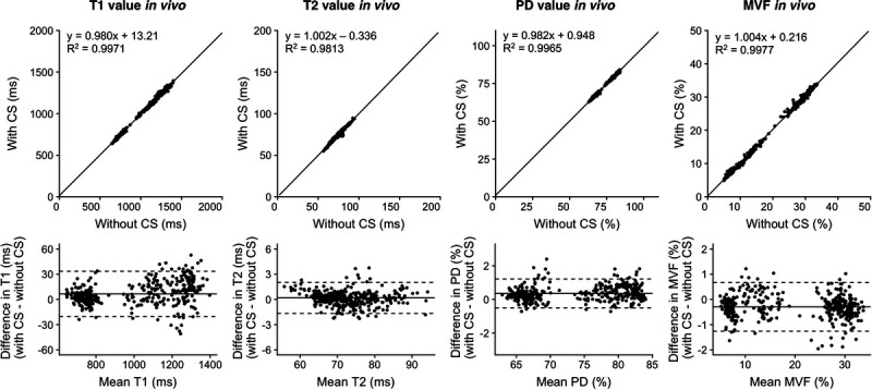
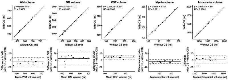
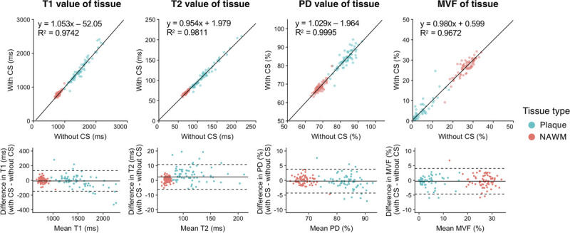
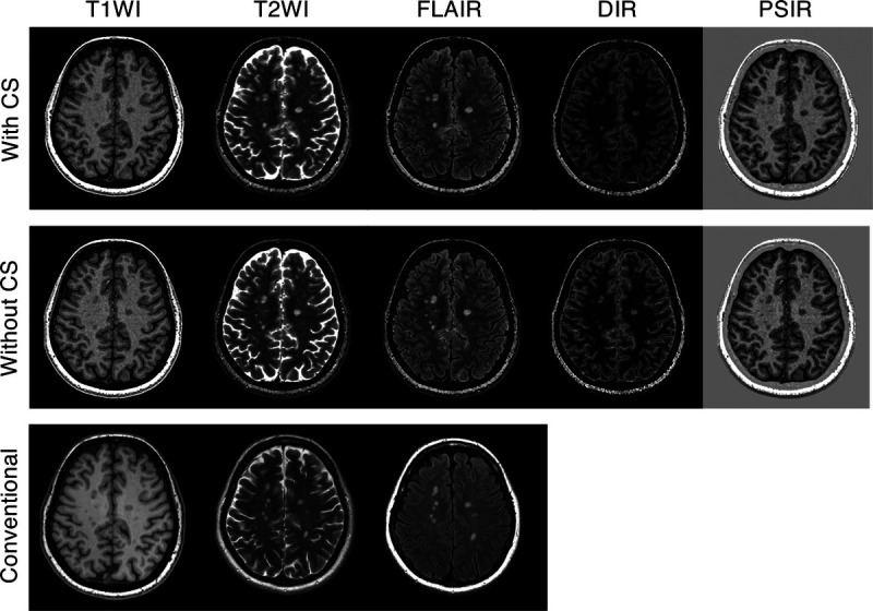
The linearity of both phantom and volunteer measurements in T1, T2, and PD values obtained with and without CS was very strong (R2 = 0.9901-1.000). The tissue segmentation obtained with and without CS also had high linearity (R2 = 0.987-0.999). The quantitative tissue values of the plaques and NAWM obtained with CS showed high linearity with those without CS (R2 = 0.967-1.000). There were no significant differences in overall image quality between synthetic contrast-weighted images obtained with and without CS (P = 0.17-0.99).
Multiparametric imaging of the whole brain based on 3D-QALAS can be accelerated using CS while preserving tissue quantitative values, tissue segmentation, and quality of synthetic images.
本研究旨在开发一种基于 3D 定量的加速磁共振成像方法,使用带有 T2 准备脉冲的交错 Look-Locker 采集序列(3D-QALAS)结合压缩感知(CS),并评估 CS 对定量映射、组织分割和合成图像质量的影响。
磁共振成像系统体模,包含多个具有标准化 T1、T2 和质子密度(PD)值的腔室;10 名健康志愿者;12 名多发性硬化症患者。使用 3D-QALAS 序列(有和无 CS)和常规对比加权成像对体模、志愿者和患者进行扫描。3D-QALAS 序列有和无 CS 的扫描时间分别为 5:56 和 11:11。对于健康志愿者,基于测量的 T1、T2 和 PD 进行脑容积测量和髓鞘估计。对于多发性硬化症患者,测量斑块和对侧正常外观白质(NAWM)中的平均 T1、T2、PD 和髓鞘量。对有和无 CS 的数据集获得的每个指标进行简单线性回归分析和 Bland-Altman 分析。为了比较合成和常规对比加权图像的整体图像质量和结构描绘,由 2 名神经放射科医生以盲法进行病例对照随机阅读。
有和无 CS 的体模和志愿者 T1、T2 和 PD 值的测量线性非常强(R2=0.9901-1.000)。有和无 CS 的组织分割也具有高线性(R2=0.987-0.999)。有和无 CS 的斑块和 NAWM 的定量组织值具有高线性(R2=0.967-1.000)。有和无 CS 的合成对比加权图像的整体图像质量没有显著差异(P=0.17-0.99)。
基于 3D-QALAS 的全脑多参数成像可以使用 CS 加速,同时保留组织定量值、组织分割和合成图像质量。