• 文献检索
  • 文档翻译
  • 深度研究
  • 学术资讯
  • Suppr Zotero 插件Zotero 插件
  • 邀请有礼
  • 套餐&价格
  • 历史记录
应用&插件
Suppr Zotero 插件Zotero 插件浏览器插件Mac 客户端Windows 客户端微信小程序
定价
高级版会员购买积分包购买API积分包
服务
文献检索文档翻译深度研究API 文档MCP 服务
关于我们
关于 Suppr公司介绍联系我们用户协议隐私条款
关注我们

Suppr 超能文献

核心技术专利:CN118964589B侵权必究
粤ICP备2023148730 号-1Suppr @ 2026

文献检索

告别复杂PubMed语法,用中文像聊天一样搜索,搜遍4000万医学文献。AI智能推荐,让科研检索更轻松。

立即免费搜索

文件翻译

保留排版,准确专业,支持PDF/Word/PPT等文件格式,支持 12+语言互译。

免费翻译文档

深度研究

AI帮你快速写综述,25分钟生成高质量综述,智能提取关键信息,辅助科研写作。

立即免费体验

使用3D-QALAS在一次屏气过程中同步进行三维心肌T1和T2映射。

Simultaneous three-dimensional myocardial T1 and T2 mapping in one breath hold with 3D-QALAS.

作者信息

Kvernby Sofia, Warntjes Marcel Jan Bertus, Haraldsson Henrik, Carlhäll Carl-Johan, Engvall Jan, Ebbers Tino

机构信息

Division of Cardiovascular Medicine, Department of Medical and Health Sciences, Linköping University, Linköping, Sweden.

出版信息

J Cardiovasc Magn Reson. 2014 Dec 20;16(1):102. doi: 10.1186/s12968-014-0102-0.

DOI:10.1186/s12968-014-0102-0
PMID:25526880
原文链接:https://pmc.ncbi.nlm.nih.gov/articles/PMC4272556/
Abstract

BACKGROUND

Quantification of the longitudinal- and transverse relaxation time in the myocardium has shown to provide important information in cardiac diagnostics. Methods for cardiac relaxation time mapping generally demand a long breath hold to measure either T1 or T2 in a single 2D slice. In this paper we present and evaluate a novel method for 3D interleaved T1 and T2 mapping of the whole left ventricular myocardium within a single breath hold of 15 heartbeats.

METHODS

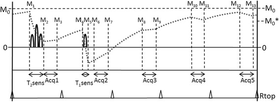
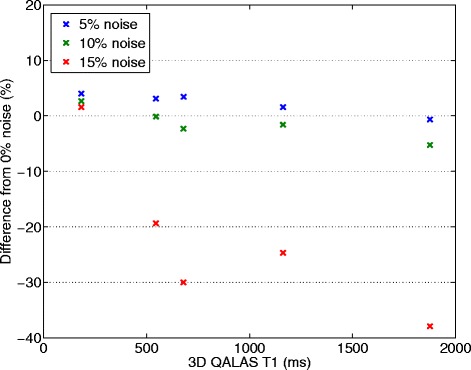
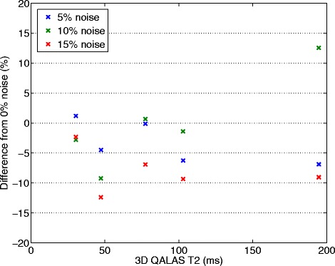
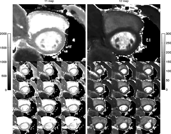
The 3D-QALAS (3D-quantification using an interleaved Look-Locker acquisition sequence with T2 preparation pulse) is based on a 3D spoiled Turbo Field Echo sequence using inversion recovery with interleaved T2 preparation. Quantification of both T1 and T2 in a volume of 13 slices with a resolution of 2.0x2.0x6.0 mm is obtained from five measurements by using simulations of the longitudinal magnetizations Mz. This acquisition scheme is repeated three times to sample k-space. The method was evaluated both in-vitro (validated against Inversion Recovery and Multi Echo) and in-vivo (validated against MOLLI and Dual Echo).

RESULTS

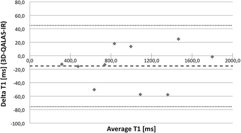
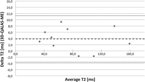
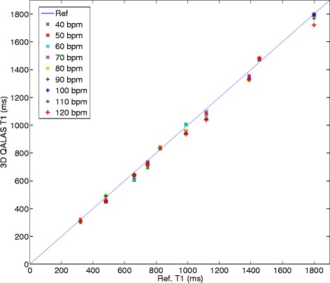
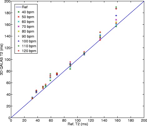
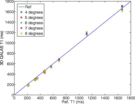
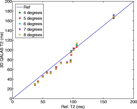
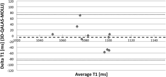
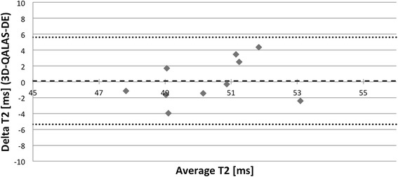
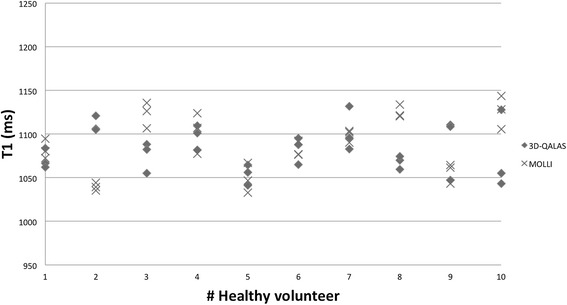
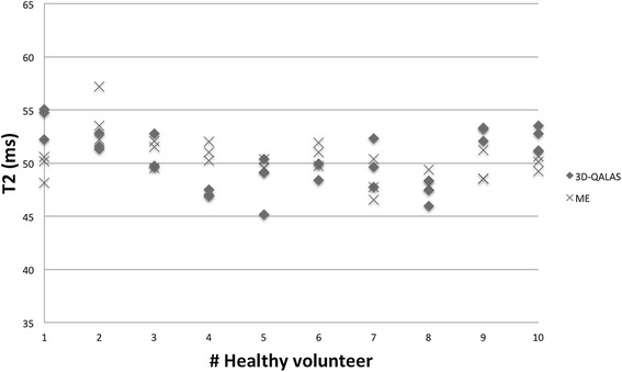
In-vitro, a strong relation was found between 3D-QALAS and Inversion Recovery (R = 0.998; N = 10; p < 0.01) and between 3D-QALAS and Multi Echo (R = 0.996; N = 10; p < 0.01). The 3D-QALAS method showed no dependence on e.g. heart rate in the interval of 40-120 bpm. In healthy myocardium, the mean T1 value was 1083 ± 43 ms (mean ± SD) for 3D-QALAS and 1089 ± 54 ms for MOLLI, while the mean T2 value was 50.4 ± 3.6 ms 3D-QALAS and 50.3 ± 3.5 ms for Dual Echo. No significant difference in in-vivo relaxation times was found between 3D-QALAS and MOLLI (N = 10; p = 0.65) respectively 3D-QALAS and Dual Echo (N = 10; p = 0.925) for the ten healthy volunteers.

CONCLUSIONS

The 3D-QALAS method has demonstrated good accuracy and intra-scan variability both in-vitro and in-vivo. It allows rapid acquisition and provides quantitative information of both T1 and T2 relaxation times in the same scan with full coverage of the left ventricle, enabling clinical application in a broader spectrum of cardiac disorders.

摘要

背景

心肌纵向和横向弛豫时间的定量分析已被证明能为心脏诊断提供重要信息。心脏弛豫时间映射方法通常需要长时间屏气以在单个二维切片中测量T1或T2。在本文中,我们提出并评估了一种新方法,可在15次心跳的单次屏气内对整个左心室心肌进行三维交错T1和T2映射。

方法

三维QALAS(使用带有T2准备脉冲的交错Look-Locker采集序列进行三维定量)基于一种三维扰相梯度回波序列,采用反转恢复和交错T2准备。通过对纵向磁化强度Mz进行模拟,从五次测量中获得13个切片、分辨率为2.0×2.0×6.0毫米的体积内T1和T2的定量结果。该采集方案重复三次以采样k空间。该方法在体外(与反转恢复和多回波进行验证)和体内(与MOLLI和双回波进行验证)均进行了评估。

结果

在体外,三维QALAS与反转恢复之间(R = 0.998;N = 10;p < 0.01)以及三维QALAS与多回波之间(R = 0.996;N = 10;p < 0.01)发现有很强的相关性。三维QALAS方法在40 - 120次/分钟的心率区间内不依赖于例如心率。在健康心肌中,三维QALAS的平均T1值为1083±43毫秒(平均值±标准差),MOLLI为1089±54毫秒,而三维QALAS的平均T2值为50.4±3.6毫秒,双回波为50.3±3.5毫秒。对于十名健康志愿者,三维QALAS与MOLLI(N = 10;p = 0.65)以及三维QALAS与双回波(N = 10;p = 0.925)之间在体内弛豫时间上未发现显著差异。

结论

三维QALAS方法在体外和体内均显示出良好的准确性和扫描内变异性。它允许快速采集,并在同一扫描中提供T1和T2弛豫时间的定量信息,且能完全覆盖左心室,从而能够在更广泛的心脏疾病中得到临床应用。

https://cdn.ncbi.nlm.nih.gov/pmc/blobs/933f/4272556/fafb2bdb09b4/12968_2014_102_Fig15_HTML.jpg
https://cdn.ncbi.nlm.nih.gov/pmc/blobs/933f/4272556/01d7887cee63/12968_2014_102_Fig1_HTML.jpg
https://cdn.ncbi.nlm.nih.gov/pmc/blobs/933f/4272556/29c5ce2adeb5/12968_2014_102_Fig2_HTML.jpg
https://cdn.ncbi.nlm.nih.gov/pmc/blobs/933f/4272556/91ab3d2c6308/12968_2014_102_Fig3_HTML.jpg
https://cdn.ncbi.nlm.nih.gov/pmc/blobs/933f/4272556/7688d77a532a/12968_2014_102_Fig4_HTML.jpg
https://cdn.ncbi.nlm.nih.gov/pmc/blobs/933f/4272556/e602c0cd9bbf/12968_2014_102_Fig5_HTML.jpg
https://cdn.ncbi.nlm.nih.gov/pmc/blobs/933f/4272556/c64d1d9314af/12968_2014_102_Fig6_HTML.jpg
https://cdn.ncbi.nlm.nih.gov/pmc/blobs/933f/4272556/295254fe233e/12968_2014_102_Fig7_HTML.jpg
https://cdn.ncbi.nlm.nih.gov/pmc/blobs/933f/4272556/ef36d60188f0/12968_2014_102_Fig8_HTML.jpg
https://cdn.ncbi.nlm.nih.gov/pmc/blobs/933f/4272556/cf8acf19309b/12968_2014_102_Fig9_HTML.jpg
https://cdn.ncbi.nlm.nih.gov/pmc/blobs/933f/4272556/9ce423cec284/12968_2014_102_Fig10_HTML.jpg
https://cdn.ncbi.nlm.nih.gov/pmc/blobs/933f/4272556/015b95056d74/12968_2014_102_Fig11_HTML.jpg
https://cdn.ncbi.nlm.nih.gov/pmc/blobs/933f/4272556/b86fb0c854f8/12968_2014_102_Fig12_HTML.jpg
https://cdn.ncbi.nlm.nih.gov/pmc/blobs/933f/4272556/bb4293a1228c/12968_2014_102_Fig13_HTML.jpg
https://cdn.ncbi.nlm.nih.gov/pmc/blobs/933f/4272556/bff3d25b782a/12968_2014_102_Fig14_HTML.jpg
https://cdn.ncbi.nlm.nih.gov/pmc/blobs/933f/4272556/fafb2bdb09b4/12968_2014_102_Fig15_HTML.jpg

相似文献

1
Simultaneous three-dimensional myocardial T1 and T2 mapping in one breath hold with 3D-QALAS.使用3D-QALAS在一次屏气过程中同步进行三维心肌T1和T2映射。
J Cardiovasc Magn Reson. 2014 Dec 20;16(1):102. doi: 10.1186/s12968-014-0102-0.
2
Clinical feasibility of 3D-QALAS - Single breath-hold 3D myocardial T1- and T2-mapping.3D-QALAS的临床可行性——单次屏气3D心肌T1和T2映射
Magn Reson Imaging. 2017 May;38:13-20. doi: 10.1016/j.mri.2016.12.014. Epub 2016 Dec 18.
3
Liver T1 Relaxation Quantification Using a 3-Dimensional Interleaved Look-Locker Acquisition With T2 Preparation Pulse Sequence (3D-QALAS): Comparison With Conventional 2-Dimensional MOLLI.使用带有T2准备脉冲序列的三维交错锁相环采集技术(3D-QALAS)进行肝脏T1弛豫定量分析:与传统二维MOLLI的比较
J Comput Assist Tomogr. 2023;47(3):350-354. doi: 10.1097/RCT.0000000000001409. Epub 2022 Dec 27.
4
Three-dimensional high-resolution simultaneous quantitative mapping of the whole brain with 3D-QALAS: An accuracy and repeatability study.三维高分辨率全脑同时定量图谱成像(3D-QALAS):一项准确性和可重复性研究。
Magn Reson Imaging. 2019 Nov;63:235-243. doi: 10.1016/j.mri.2019.08.031. Epub 2019 Aug 21.
5
[Evaluation of the Accuracy of Relaxation Time Measurements Using 3D-QALAS at 3.0 T MRI and Comparison with 2D-MDME].[使用3.0T磁共振成像的3D-QALAS评估弛豫时间测量的准确性并与2D-MDME进行比较]
Nihon Hoshasen Gijutsu Gakkai Zasshi. 2023 Jul 20;79(7):663-673. doi: 10.6009/jjrt.2023-1343. Epub 2023 May 19.
6
Shortened Modified Look-Locker Inversion recovery (ShMOLLI) for clinical myocardial T1-mapping at 1.5 and 3 T within a 9 heartbeat breathhold.1.5T 和 3T 临床心肌 T1 映射的缩短改良 Look-Locker 反转恢复(ShMOLLI),在 9 次心跳屏气内完成。
J Cardiovasc Magn Reson. 2010 Nov 19;12(1):69. doi: 10.1186/1532-429X-12-69.
7
Simultaneous T1 and T2 quantification of the myocardium using cardiac balanced-SSFP inversion recovery with interleaved sampling acquisition (CABIRIA).使用具有交错采样采集的心脏平衡稳态自由进动反转恢复序列(CABIRIA)对心肌进行T1和T2同时定量分析。
Magn Reson Med. 2015 Aug;74(2):365-71. doi: 10.1002/mrm.25402. Epub 2014 Aug 11.
8
Free-running simultaneous myocardial T1/T2 mapping and cine imaging with 3D whole-heart coverage and isotropic spatial resolution.自由运行的同时心肌 T1/T2 映射和电影成像,具有 3D 全心覆盖和各向同性空间分辨率。
Magn Reson Imaging. 2019 Nov;63:159-169. doi: 10.1016/j.mri.2019.08.008. Epub 2019 Aug 16.
9
Myocardial T-mapping at 3T using saturation-recovery: reference values, precision and comparison with MOLLI.使用饱和恢复法在3T下进行心肌T值映射:参考值、精密度及与MOLLI的比较
J Cardiovasc Magn Reson. 2016 Nov 18;18(1):84. doi: 10.1186/s12968-016-0302-x.
10
Time-efficient, high-resolution 3T whole-brain relaxometry using 3D-QALAS with wave-CAIPI readouts.采用 3D-QALAS 与波 CAIPI 读出的高效、高分辨率 3T 全脑 relaxometry。
Magn Reson Med. 2024 Feb;91(2):630-639. doi: 10.1002/mrm.29865. Epub 2023 Sep 14.

引用本文的文献

1
RF phase modulation improves quantitative transient state sequences under constrained conditions.射频相位调制可改善受限条件下的定量瞬态序列。
MAGMA. 2025 Sep 11. doi: 10.1007/s10334-025-01293-9.
2
Adaptive Radiation Therapy for Head and Neck Cancer.头颈部癌的自适应放射治疗
ArXiv. 2025 Aug 1:arXiv:2508.00651v1.
3
Enhanced Partial Fourier MRI With Zero-Shot Deep Untrained Priors.基于零样本深度未训练先验的增强部分傅里叶磁共振成像

本文引用的文献

1
T1-mapping in the heart: accuracy and precision.心脏 T1-mapping:准确性和精密度。
J Cardiovasc Magn Reson. 2014 Jan 4;16(1):2. doi: 10.1186/1532-429X-16-2.
2
Myocardial T1 mapping and extracellular volume quantification: a Society for Cardiovascular Magnetic Resonance (SCMR) and CMR Working Group of the European Society of Cardiology consensus statement.心肌 T1 映射和细胞外容积定量:心血管磁共振学会 (SCMR) 和欧洲心脏病学会 CMR 工作组的共识声明。
J Cardiovasc Magn Reson. 2013 Oct 14;15(1):92. doi: 10.1186/1532-429X-15-92.
3
Standardization of T1 measurements with MOLLI in differentiation between health and disease--the ConSept study.
IEEE Access. 2024;12:182157-182170. doi: 10.1109/access.2024.3508761. Epub 2024 Nov 29.
4
Technical development and In Silico implementation of SyntheticMR in head and neck adaptive radiation therapy: A prospective R-IDEAL stage 0/1 technology development report.合成磁共振成像在头颈部自适应放射治疗中的技术开发与计算机模拟实现:一项前瞻性R-IDEAL 0/1期技术开发报告
J Appl Clin Med Phys. 2025 Jul;26(7):e70134. doi: 10.1002/acm2.70134. Epub 2025 Jul 11.
5
Vendor-agnostic 3D multiparametric relaxometry improves cross-platform reproducibility.与供应商无关的3D多参数弛豫测量法提高了跨平台的可重复性。
Magn Reson Med. 2025 Sep;94(3):937-948. doi: 10.1002/mrm.30566. Epub 2025 May 26.
6
Neuroimaging Reader Study on Clinical Sensitivity and Specificity Using Synthetic MRI Based on MR Quantification.基于磁共振定量的合成磁共振成像在临床敏感性和特异性方面的神经影像学读者研究
AJNR Am J Neuroradiol. 2025 Jun 3;46(6):1196-1202. doi: 10.3174/ajnr.A8631.
7
Comparison of test-retest repeatability of DESPOT and 3D-QALAS for T and T mapping.DESPOT和3D-QALAS用于T和T映射的重测重复性比较。
Quant Imaging Med Surg. 2025 May 1;15(5):3807-3823. doi: 10.21037/qims-24-1870. Epub 2025 Apr 24.
8
Model-based deep learning with fully connected neural networks for accelerated magnetic resonance parameter mapping.基于模型的全连接神经网络深度学习用于加速磁共振参数映射
Int J Comput Assist Radiol Surg. 2025 May 3. doi: 10.1007/s11548-025-03356-7.
9
Technical Optimization of SyntheticMR for the Head and Neck on a 3T MR-Simulator and 1.5T MR-Linac: A Prospective R-IDEAL Stage 2a Technology Innovation Report.3T磁共振模拟机和1.5T磁共振直线加速器上用于头颈部位的合成磁共振技术优化:一项前瞻性R-IDEAL 2a期技术创新报告。
medRxiv. 2025 Apr 10:2025.04.08.25325491. doi: 10.1101/2025.04.08.25325491.
10
Evaluation of a Novel Quantitative Multiparametric MR Sequence for Radiation Therapy Treatment Response Assessment.一种用于放射治疗反应评估的新型定量多参数磁共振序列的评估
ArXiv. 2025 Mar 28:arXiv:2503.22640v1.
MOLLI 测量 T1 值在健康与疾病鉴别诊断中的标准化——ConSept 研究。
J Cardiovasc Magn Reson. 2013 Sep 11;15(1):78. doi: 10.1186/1532-429X-15-78.
4
Myocardial T1 and T2 mapping at 3 T: reference values, influencing factors and implications.3T下的心肌T1和T2成像:参考值、影响因素及意义
J Cardiovasc Magn Reson. 2013 Jun 18;15(1):53. doi: 10.1186/1532-429X-15-53.
5
Non-contrast T1-mapping detects acute myocardial edema with high diagnostic accuracy: a comparison to T2-weighted cardiovascular magnetic resonance.非对比 T1 映射以高诊断准确性检测急性心肌水肿:与 T2 加权心血管磁共振的比较。
J Cardiovasc Magn Reson. 2012 Jun 21;14(1):42. doi: 10.1186/1532-429X-14-42.
6
Myocardial edema as detected by pre-contrast T1 and T2 CMR delineates area at risk associated with acute myocardial infarction.心肌水肿在对比前 T1 和 T2 CMR 上可检测到,可划定与急性心肌梗死相关的危险区域。
JACC Cardiovasc Imaging. 2012 Jun;5(6):596-603. doi: 10.1016/j.jcmg.2012.01.016.
7
Multiple 3D inversion recovery imaging for volume T1 mapping of the heart.多层面 3D 反转恢复成像用于心脏容积 T1 mapping。
Magn Reson Med. 2013 Jan;69(1):163-70. doi: 10.1002/mrm.24248. Epub 2012 Apr 5.
8
Myocardial T1 and extracellular volume fraction mapping at 3 tesla.3T 下的心肌 T1 和细胞外容积分数测绘
J Cardiovasc Magn Reson. 2011 Nov 28;13(1):75. doi: 10.1186/1532-429X-13-75.
9
Direct T2 quantification of myocardial edema in acute ischemic injury.直接 T2 定量评估急性缺血性损伤中的心肌水肿。
JACC Cardiovasc Imaging. 2011 Mar;4(3):269-78. doi: 10.1016/j.jcmg.2010.09.023.
10
Shortened Modified Look-Locker Inversion recovery (ShMOLLI) for clinical myocardial T1-mapping at 1.5 and 3 T within a 9 heartbeat breathhold.1.5T 和 3T 临床心肌 T1 映射的缩短改良 Look-Locker 反转恢复(ShMOLLI),在 9 次心跳屏气内完成。
J Cardiovasc Magn Reson. 2010 Nov 19;12(1):69. doi: 10.1186/1532-429X-12-69.