• 文献检索
  • 文档翻译
  • 深度研究
  • 学术资讯
  • Suppr Zotero 插件Zotero 插件
  • 邀请有礼
  • 套餐&价格
  • 历史记录
应用&插件
Suppr Zotero 插件Zotero 插件浏览器插件Mac 客户端Windows 客户端微信小程序
定价
高级版会员购买积分包购买API积分包
服务
文献检索文档翻译深度研究API 文档MCP 服务
关于我们
关于 Suppr公司介绍联系我们用户协议隐私条款
关注我们

Suppr 超能文献

核心技术专利:CN118964589B侵权必究
粤ICP备2023148730 号-1Suppr @ 2026

文献检索

告别复杂PubMed语法,用中文像聊天一样搜索,搜遍4000万医学文献。AI智能推荐,让科研检索更轻松。

立即免费搜索

文件翻译

保留排版,准确专业,支持PDF/Word/PPT等文件格式,支持 12+语言互译。

免费翻译文档

深度研究

AI帮你快速写综述,25分钟生成高质量综述,智能提取关键信息,辅助科研写作。

立即免费体验

MK-Curve 提高了对精神病临床高危人群白质改变的敏感性。

MK-Curve improves sensitivity to identify white matter alterations in clinical high risk for psychosis.

机构信息

Department of Radiology, Brigham and Women's Hospital, Harvard Medical School, Boston, MA, USA.

Department of Psychiatry, Brigham and Women's Hospital, Harvard Medical School, Boston, MA, USA.

出版信息

Neuroimage. 2021 Feb 1;226:117564. doi: 10.1016/j.neuroimage.2020.117564. Epub 2020 Dec 4.

DOI:10.1016/j.neuroimage.2020.117564
PMID:33285331
原文链接:https://pmc.ncbi.nlm.nih.gov/articles/PMC7873589/
Abstract

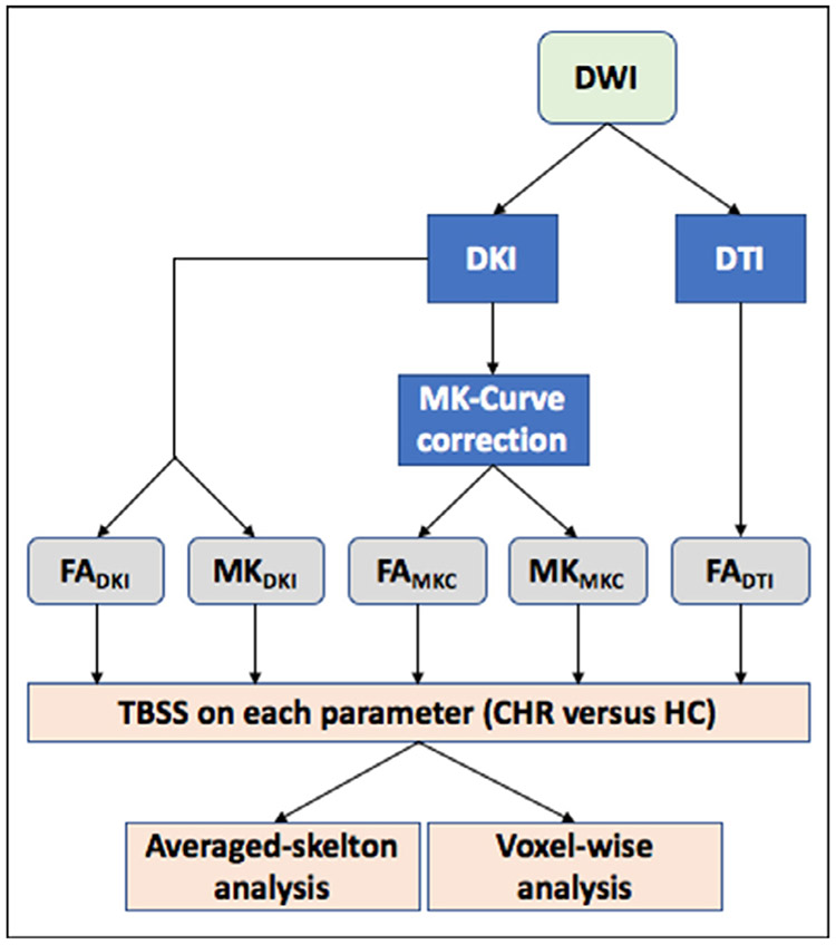
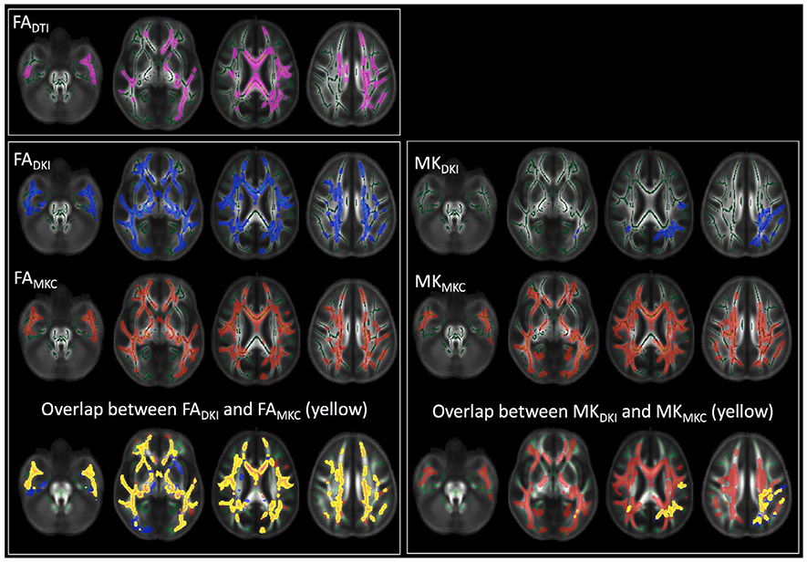
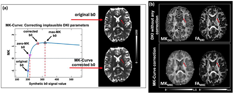
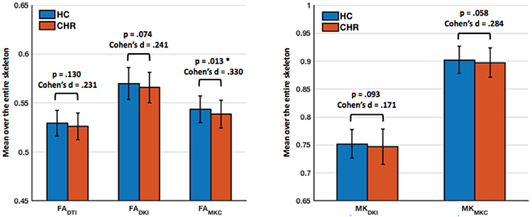
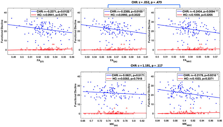
Diffusion kurtosis imaging (DKI) is a diffusion MRI approach that enables the measurement of brain microstructural properties, reflecting molecular restrictions and tissue heterogeneity. DKI parameters such as mean kurtosis (MK) provide additional subtle information to that provided by popular diffusion tensor imaging (DTI) parameters, and thus have been considered useful to detect white matter abnormalities, especially in populations that are not expected to show severe brain pathologies. However, DKI parameters often yield artifactual output values that are outside of the biologically plausible range, which diminish sensitivity to identify true microstructural changes. Recently we have proposed the mean-kurtosis-curve (MK-Curve) method to correct voxels with implausible DKI parameters, and demonstrated its improved performance against other approaches that correct artifacts in DKI. In this work, we aimed to evaluate the utility of the MK-Curve method to improve the identification of white matter abnormalities in group comparisons. To do so, we compared group differences, with and without the MK-Curve correction, between 115 individuals at clinical high risk for psychosis (CHR) and 93 healthy controls (HCs). We also compared the correlation of the corrected and uncorrected DKI parameters with clinical characteristics. Following the MK-curve correction, the group differences had larger effect sizes and higher statistical significance (i.e., lower p-values), demonstrating increased sensitivity to detect group differences, in particular in MK. Furthermore, the MK-curve-corrected DKI parameters displayed stronger correlations with clinical variables in CHR individuals, demonstrating the clinical relevance of the corrected parameters. Overall, following the MK-curve correction our analyses found widespread lower MK in CHR that overlapped with lower fractional anisotropy (FA), and both measures were significantly correlated with a decline in functioning and with more severe symptoms. These observations further characterize white matter alterations in the CHR stage, demonstrating that MK and FA abnormalities are widespread, and mostly overlap. The improvement in group differences and stronger correlation with clinical variables suggest that applying MK-curve would be beneficial for the detection and characterization of subtle group differences in other experiments as well.

摘要

扩散峰度成像(DKI)是一种扩散 MRI 方法,可用于测量脑微观结构特性,反映分子限制和组织异质性。DKI 参数(如平均峰度(MK))提供了比流行的扩散张量成像(DTI)参数更多的细微信息,因此被认为有助于检测白质异常,尤其是在那些预计不会出现严重脑病变的人群中。然而,DKI 参数通常会产生超出生物学合理范围的人为输出值,从而降低了识别真正微观结构变化的敏感性。最近,我们提出了平均峰度曲线(MK-Curve)方法来校正不合理的 DKI 参数的体素,并证明了它在纠正 DKI 伪影方面优于其他方法的性能。在这项工作中,我们旨在评估 MK-Curve 方法在组间比较中改善白质异常识别的效用。为此,我们比较了 115 名临床高风险精神分裂症(CHR)个体和 93 名健康对照(HC)个体的组间差异,分别有无 MK-Curve 校正。我们还比较了校正和未校正的 DKI 参数与临床特征的相关性。在进行 MK-Curve 校正后,组间差异的效应大小更大,统计学意义更高(即 p 值更低),表明校正后对组间差异的检测敏感性更高,尤其是在 MK 上。此外,在 CHR 个体中,MK-curve 校正后的 DKI 参数与临床变量的相关性更强,表明校正后的参数具有临床相关性。总体而言,在进行 MK-Curve 校正后,我们的分析发现 CHR 患者的 MK 普遍较低,与 FA 降低重叠,并且这两个指标都与功能下降和更严重的症状显著相关。这些观察结果进一步描述了 CHR 阶段的白质改变,表明 MK 和 FA 异常是广泛存在的,并且大部分重叠。组间差异的改善和与临床变量的更强相关性表明,在其他实验中应用 MK-Curve 将有助于检测和描述细微的组间差异。

https://cdn.ncbi.nlm.nih.gov/pmc/blobs/c80e/7873589/11f314bc2aa3/nihms-1666837-f0005.jpg
https://cdn.ncbi.nlm.nih.gov/pmc/blobs/c80e/7873589/aa128e7aaf05/nihms-1666837-f0001.jpg
https://cdn.ncbi.nlm.nih.gov/pmc/blobs/c80e/7873589/2be6037e85df/nihms-1666837-f0002.jpg
https://cdn.ncbi.nlm.nih.gov/pmc/blobs/c80e/7873589/2042467bd82a/nihms-1666837-f0003.jpg
https://cdn.ncbi.nlm.nih.gov/pmc/blobs/c80e/7873589/04b666496396/nihms-1666837-f0004.jpg
https://cdn.ncbi.nlm.nih.gov/pmc/blobs/c80e/7873589/11f314bc2aa3/nihms-1666837-f0005.jpg

相似文献

1
MK-Curve improves sensitivity to identify white matter alterations in clinical high risk for psychosis.MK-Curve 提高了对精神病临床高危人群白质改变的敏感性。
Neuroimage. 2021 Feb 1;226:117564. doi: 10.1016/j.neuroimage.2020.117564. Epub 2020 Dec 4.
2
MK-curve - Characterizing the relation between mean kurtosis and alterations in the diffusion MRI signal.MK 曲线——刻画平均峰度与扩散磁共振信号改变之间的关系。
Neuroimage. 2019 Aug 1;196:68-80. doi: 10.1016/j.neuroimage.2019.04.015. Epub 2019 Apr 10.
3
Mean kurtosis-Curve (MK-Curve) correction improves the test-retest reproducibility of diffusion kurtosis imaging at 3 T.平均峰度曲线(MK-Curve)校正可提高 3T 下扩散峰度成像的复测可重复性。
NMR Biomed. 2023 Mar;36(3):e4856. doi: 10.1002/nbm.4856. Epub 2022 Nov 22.
4
Evaluation of patients with relapsing-remitting multiple sclerosis using tract-based spatial statistics analysis: diffusion kurtosis imaging.使用基于纤维束的空间统计学分析评估复发缓解型多发性硬化症患者:扩散峰度成像
BMC Neurol. 2018 Aug 7;18(1):108. doi: 10.1186/s12883-018-1108-2.
5
Differences in Gaussian diffusion tensor imaging and non-Gaussian diffusion kurtosis imaging model-based estimates of diffusion tensor invariants in the human brain.基于高斯扩散张量成像和非高斯扩散峰度成像模型的人脑扩散张量不变量估计差异。
Med Phys. 2016 May;43(5):2464. doi: 10.1118/1.4946819.
6
Microstructural white matter abnormalities in overactive bladder syndrome evaluation with diffusion kurtosis imaging tract-based spatial statistics analysis.应用扩散峰度成像基于体素的空间统计学分析评估膀胱过度活动症的脑微观结构白质异常。
World J Urol. 2024 Jan 13;42(1):36. doi: 10.1007/s00345-023-04709-0.
7
Performances of diffusion kurtosis imaging and diffusion tensor imaging in detecting white matter abnormality in schizophrenia.扩散峰度成像和扩散张量成像在检测精神分裂症白质异常中的表现。
Neuroimage Clin. 2014 Dec 9;7:170-6. doi: 10.1016/j.nicl.2014.12.008. eCollection 2015.
8
Comparative Evaluation of Diffusion Kurtosis Imaging and Diffusion Tensor Imaging in Detecting Cerebral Microstructural Changes in Alzheimer Disease.比较扩散峰度成像和扩散张量成像在阿尔茨海默病脑微结构变化检测中的应用。
Acad Radiol. 2022 Mar;29 Suppl 3:S63-S70. doi: 10.1016/j.acra.2021.01.018. Epub 2021 Feb 18.
9
Microstructural alterations in different types of lesions and their perilesional white matter in relapsing-remitting multiple sclerosis based on diffusion kurtosis imaging.基于扩散峰度成像的复发缓解型多发性硬化症不同类型病灶及其病灶周围白质的微观结构改变。
Mult Scler Relat Disord. 2023 Mar;71:104572. doi: 10.1016/j.msard.2023.104572. Epub 2023 Feb 19.
10
Diffusion tensor and kurtosis imaging reveal microstructural changes in the trigeminal nerves of patients with trigeminal neuralgia.弥散张量和峰度成像显示三叉神经痛患者三叉神经的结构变化。
Eur Radiol. 2023 Nov;33(11):8046-8054. doi: 10.1007/s00330-023-09762-2. Epub 2023 May 31.

引用本文的文献

1
Exploring mean kurtosis in MR diffusion kurtosis imaging for early detection of lumbar spine degeneration: a systematic review.探索磁共振扩散峰度成像中的平均峰度用于早期检测腰椎退变:一项系统综述
F1000Res. 2025 Apr 15;14:440. doi: 10.12688/f1000research.163638.1. eCollection 2025.
2
White matter microstructure alterations in early psychosis and schizophrenia.早期精神病和精神分裂症中的白质微观结构改变
Transl Psychiatry. 2025 May 23;15(1):179. doi: 10.1038/s41398-025-03397-1.
3
A narrative literature review of white matter microstructure in individuals at clinical high risk for psychosis.

本文引用的文献

1
Deep learning based segmentation of brain tissue from diffusion MRI.基于深度学习的弥散磁共振成像脑组织结构分割。
Neuroimage. 2021 Jun;233:117934. doi: 10.1016/j.neuroimage.2021.117934. Epub 2021 Mar 16.
2
Progression from being at-risk to psychosis: next steps.从高危状态发展到精神病:后续步骤。
NPJ Schizophr. 2020 Oct 5;6(1):27. doi: 10.1038/s41537-020-00117-0.
3
White Matter Microstructure across the Psychosis Spectrum.精神病谱系中的白质微观结构
对临床高危精神病个体白质微观结构的叙述性文献综述。
Psychoradiology. 2024 Dec 24;5:kkae031. doi: 10.1093/psyrad/kkae031. eCollection 2025.
4
The MR neuroimaging protocol for the Accelerating Medicines Partnership® Schizophrenia Program.加速药物合作组织精神分裂症项目的磁共振神经成像方案。
Schizophrenia (Heidelb). 2025 Apr 2;11(1):52. doi: 10.1038/s41537-025-00581-6.
5
Excessive interstitial free-water in cortical gray matter preceding accelerated volume changes in individuals at clinical high risk for psychosis.皮质灰质中过多的细胞外游离水先于临床精神病高危个体的体积加速变化。
Mol Psychiatry. 2024 Nov;29(11):3623-3634. doi: 10.1038/s41380-024-02597-3. Epub 2024 Jun 3.
6
Fixel-Based Analysis Reveals Tau-Related White Matter Changes in Early Stages of Alzheimer's Disease.基于体素的分析显示,阿尔茨海默病早期阶段存在与 tau 相关的白质变化。
J Neurosci. 2024 May 1;44(18):e0538232024. doi: 10.1523/JNEUROSCI.0538-23.2024.
7
Surface-based Analyses of Diffusional Kurtosis Imaging in Amyotrophic Lateral Sclerosis: Relationship with Onset Subtypes.肌萎缩侧索硬化症中基于表面的扩散峰度成像分析:与发病亚型的关系
Magn Reson Med Sci. 2025 Jan 1;24(1):122-132. doi: 10.2463/mrms.mp.2023-0138. Epub 2024 May 29.
8
DDParcel: Deep Learning Anatomical Brain Parcellation From Diffusion MRI.DDParcel:基于弥散磁共振成像的深度学习脑区自动划分。
IEEE Trans Med Imaging. 2024 Mar;43(3):1191-1202. doi: 10.1109/TMI.2023.3331691. Epub 2024 Mar 5.
9
Microstructural Cortical Gray Matter Changes Preceding Accelerated Volume Changes in Individuals at Clinical High Risk for Psychosis.精神病临床高危个体中,微观结构皮质灰质变化先于加速的体积变化。
Res Sq. 2023 Sep 28:rs.3.rs-3179575. doi: 10.21203/rs.3.rs-3179575/v1.
10
Brain tissue segmentation in neurosurgery: a systematic analysis for quantitative tractography approaches.神经外科中的脑组织分割:用于定量束追踪方法的系统分析。
Acta Neurol Belg. 2024 Feb;124(1):1-15. doi: 10.1007/s13760-023-02170-9. Epub 2023 Jan 6.
Trends Neurosci. 2020 Jun;43(6):406-416. doi: 10.1016/j.tins.2020.03.014. Epub 2020 Apr 26.
4
SlicerDMRI: Diffusion MRI and Tractography Research Software for Brain Cancer Surgery Planning and Visualization.SlicerDMRI:用于脑癌手术规划与可视化的扩散磁共振成像及纤维束成像研究软件。
JCO Clin Cancer Inform. 2020 Mar;4:299-309. doi: 10.1200/CCI.19.00141.
5
Diffusion kurtosis imaging of gray matter in schizophrenia.精神分裂症患者脑灰质的弥散峰度成像。
Cortex. 2019 Dec;121:201-224. doi: 10.1016/j.cortex.2019.08.013. Epub 2019 Aug 29.
6
Altered Cellular White Matter But Not Extracellular Free Water on Diffusion MRI in Individuals at Clinical High Risk for Psychosis.精神分裂症临床高危个体的弥散 MRI 显示细胞外自由水无变化但细胞白质改变。
Am J Psychiatry. 2019 Oct 1;176(10):820-828. doi: 10.1176/appi.ajp.2019.18091044. Epub 2019 Jun 24.
7
MK-curve - Characterizing the relation between mean kurtosis and alterations in the diffusion MRI signal.MK 曲线——刻画平均峰度与扩散磁共振信号改变之间的关系。
Neuroimage. 2019 Aug 1;196:68-80. doi: 10.1016/j.neuroimage.2019.04.015. Epub 2019 Apr 10.
8
Test-retest reproducibility of white matter parcellation using diffusion MRI tractography fiber clustering.基于弥散磁共振成像纤维追踪的纤维聚类技术的脑白质分割的重测信度研究。
Hum Brain Mapp. 2019 Jul;40(10):3041-3057. doi: 10.1002/hbm.24579. Epub 2019 Mar 15.
9
Microstructural Changes in Higher-Order Nuclei of the Thalamus in Patients With First-Episode Psychosis.首发精神病患者丘脑高级核团的微观结构变化。
Biol Psychiatry. 2019 Jan 1;85(1):70-78. doi: 10.1016/j.biopsych.2018.05.019. Epub 2018 May 31.
10
Advances in microstructural diffusion neuroimaging for psychiatric disorders.精神障碍的微观结构扩散神经影像学进展。
Neuroimage. 2018 Nov 15;182:259-282. doi: 10.1016/j.neuroimage.2018.04.051. Epub 2018 May 2.