Univ Paris Est Creteil, Gly-CRRET, Glycobiology Cell Growth Tissue Repair and Regeneration Research Unit, Créteil, F-94010, France.
INSERM, UMR-S 1132 Bioscar, Centre Viggo Petersen, Hôpital Lariboisière, 2, Rue Ambroise Paré,, Creteil, F-94010, France.
Arthritis Res Ther. 2020 Dec 7;22(1):283. doi: 10.1186/s13075-020-02352-3.
Heparan sulfate (HS) proteoglycans (PG) may be found at the chondrocyte surface and in the pericellular cartilage matrix, and are involved in cell-cell and cell-matrix interactions. An important function of HS chains is to regulate cell fate through specific interactions with heparin-binding proteins (HBP) modulated by their complex sulfation pattern. Osteoarthritis (OA) is a joint disorder characterized by the degradation of articular cartilaginous extracellular matrix. The aim of this study was to investigate HS structure and functions in osteoarthritic cartilages compared to normal cartilages (controls).
Glycosaminoglycans (GAG) were extracted from human macroscopically normal cartilages (controls, n = 7) and (OA cartilages n = 11). HS were isolated and quantified using the DMMB quantification method. Their structure and functions were then compared using respectively a HPLC analysis and HBP binding tests and their phenotypic effects on murine chondrocytes were studied by RQ-PCR. Statistical analyzes were performed using a one-way ANOVA followed by a Dunnett's test or a t test for pairwise comparisons.
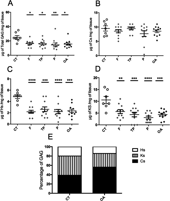
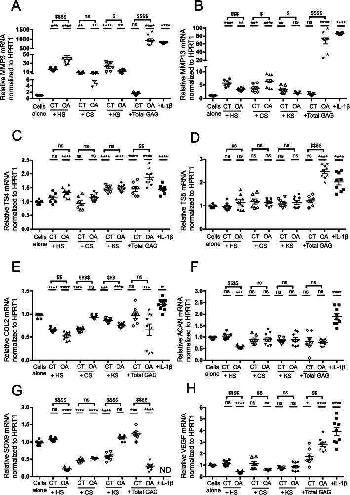
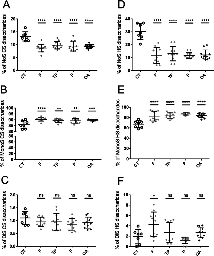
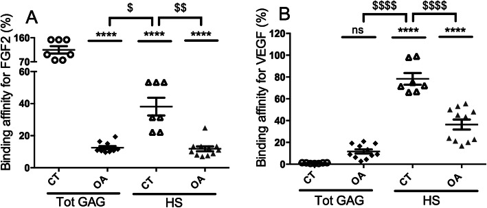
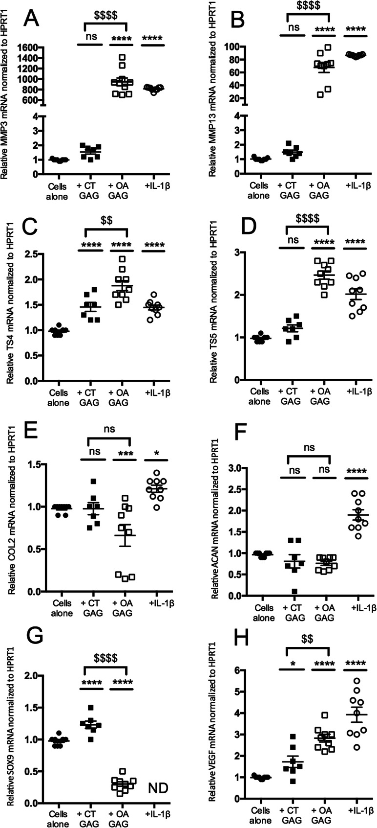
In OA, HS were characterized by increased sulfation levels compared to controls. Moreover, the capacity of these HS to bind HBP involved in the OA pathophysiological process such as FGF2 and VEGF was reduced. Chondroitin sulfates and keratan sulfates regulated these binding properties. Finally, HS from OA cartilages induced the mRNA levels of catabolic markers such as MMP3, MMP13, and TS4 and inhibited the mRNA levels of anabolic markers such as COL2, ACAN, SOX9, and VEGF in murine articular chondrocytes.
The sulfation of HS chains was increased in OA cartilages with changes in HBP binding properties and biological effects on chondrocyte phenotypes. Thus, modified HS present in altered cartilages could be a novel therapeutic target in OA.
硫酸乙酰肝素(HS)蛋白聚糖(PG)可存在于软骨细胞表面和细胞周围的软骨基质中,参与细胞-细胞和细胞-基质相互作用。HS 链的一个重要功能是通过与肝素结合蛋白(HBP)的特异性相互作用来调节细胞命运,而 HBP 的命运又受到其复杂硫酸化模式的调节。骨关节炎(OA)是一种关节疾病,其特征是关节软骨细胞外基质降解。本研究旨在比较正常软骨(对照组,n=7)和骨关节炎软骨(OA 软骨,n=11)中 HS 的结构和功能。通过 DMMB 定量法提取糖胺聚糖(GAG)。然后使用 HPLC 分析、HBP 结合试验分别比较 HS 的结构和功能,并通过 RQ-PCR 研究其对鼠软骨细胞的表型影响。使用单向方差分析(ANOVA) followed by Dunnett's test 或 t 检验进行统计学分析。
在 OA 中,与对照组相比,HS 的硫酸化水平增加。此外,这些 HS 结合 FGF2 和 VEGF 等参与 OA 病理生理过程的 HBP 的能力降低。硫酸软骨素和角质素硫酸盐调节了这些结合特性。最后,OA 软骨中的 HS 诱导了软骨细胞中分解代谢标志物如 MMP3、MMP13 和 TS4 的 mRNA 水平,并抑制了合成代谢标志物如 COL2、ACAN、SOX9 和 VEGF 的 mRNA 水平。
OA 软骨中 HS 链的硫酸化增加,HBP 结合特性和对软骨细胞表型的生物学效应发生变化。因此,改变的软骨中存在的改性 HS 可能成为 OA 的新治疗靶点。