Beck Florian, Austermann Stephanie, Bertl Kristina, Ulm Christian, Lettner Stefan, Toelly Andrea, Gahleitner André
Division of Oral Surgery, University Clinic of Dentistry, Medical University of Vienna, Vienna, Austria.
Department of Periodontology, Faculty of Odontology, University of Malmö, Malmö, Sweden.
Clin Oral Investig. 2021 Jun;25(6):3861-3871. doi: 10.1007/s00784-020-03716-4. Epub 2020 Dec 7.
To assess the reliability of judging the spatial relation between the inferior alveolar nerve (IAN) and mandibular third molar (MTM) based on MRI or CT/CBCT images.
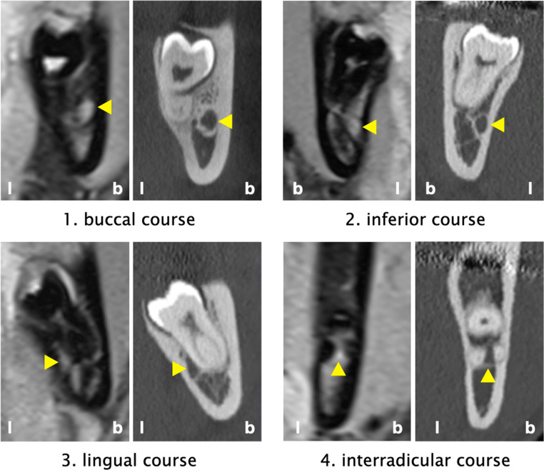
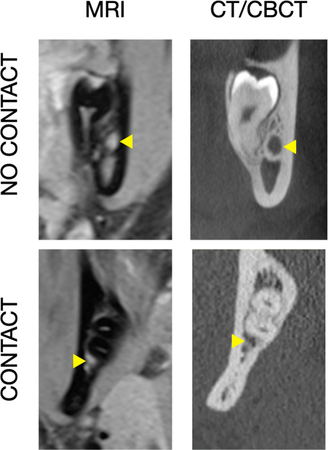
Altogether, CT/CBCT and MRI images of 87 MTMs were examined twice by 3 examiners with different degrees of experience. The course of the IAN in relation to the MTM, the presence/absence of a direct contact between IAN and MTM, and the presence of accessory IAN were determined.
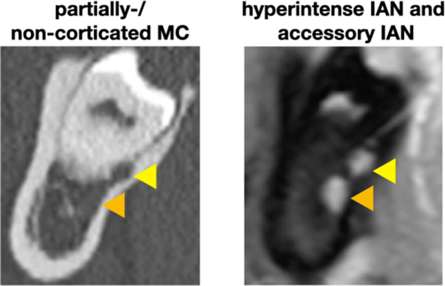
The IAN was in > 40% of the cases judged as inferior, while an interradicular position was diagnosed in < 5% of the cases. The overall agreement was good (κ = 0.72) and any disagreement between the imaging modalities was primarily among the adjacent regions, i.e., buccal/lingual/interradicular vs. inferior. CT/CBCT judgements presented a very good agreement for the inter- and intrarater comparison (κ > 0.80), while MRI judgements showed a slightly lower, but good agreement (κ = 0.74). A direct contact between IAN and MTM was diagnosed in about 65%, but in almost 20% a disagreement between the judgements based on MRI and CT/CBCT was present resulting in a moderate overall agreement (κ = 0.60). The agreement between the judgements based on MRI and CT/CBCT appeared independent of the examiner's experience and accessory IAN were described in 10 cases in MRI compared to 3 cases in CT/CBCT images.
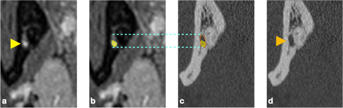
A good inter- and intrarater agreement has been observed for the assessment of the spatial relation between the IAN and MTM based on MRI images. Further, MRI images might provide advantages in the detection of accessory IAN compared to CT/CBCT.
MRI appears as viable alternative to CT/CBCT for preoperative assessment of the IAN in relation to the MTM.
基于MRI或CT/CBCT图像评估判断下牙槽神经(IAN)与下颌第三磨牙(MTM)之间空间关系的可靠性。
87颗MTM的CT/CBCT和MRI图像由3名经验程度不同的检查者检查两次。确定IAN相对于MTM的走行、IAN与MTM之间是否存在直接接触以及是否存在IAN分支。
在超过40%的病例中,IAN被判断为位于下方,而在不到5%的病例中诊断为根间位置。总体一致性良好(κ = 0.72),成像方式之间的任何差异主要存在于相邻区域,即颊侧/舌侧/根间与下方。CT/CBCT判断在不同检查者间和同一检查者内比较时一致性非常好(κ > 0.80),而MRI判断显示一致性略低,但也良好(κ = 0.74)。IAN与MTM之间的直接接触在约65%的病例中被诊断出,但在近20%的病例中,基于MRI和CT/CBCT的判断存在差异,导致总体一致性中等(κ = 0.60)。基于MRI和CT/CBCT的判断之间的一致性似乎与检查者的经验无关,MRI图像中描述了10例IAN分支,而CT/CBCT图像中为3例。
基于MRI图像评估IAN与MTM之间的空间关系时,在不同检查者间和同一检查者内均观察到良好的一致性。此外,与CT/CBCT相比,MRI图像在检测IAN分支方面可能具有优势。
对于术前评估IAN与MTM的关系,MRI似乎是CT/CBCT的可行替代方法。