Department of Diabetology and Endocrinology, Kanazawa Medical University, Uchinada, Ishikawa, Japan.
Department of Obstetrics and Gynecology, Juntendo Medical University, Bunkyo, Tokyo, Japan.
J Diabetes Investig. 2021 May;12(5):697-709. doi: 10.1111/jdi.13478. Epub 2020 Dec 31.
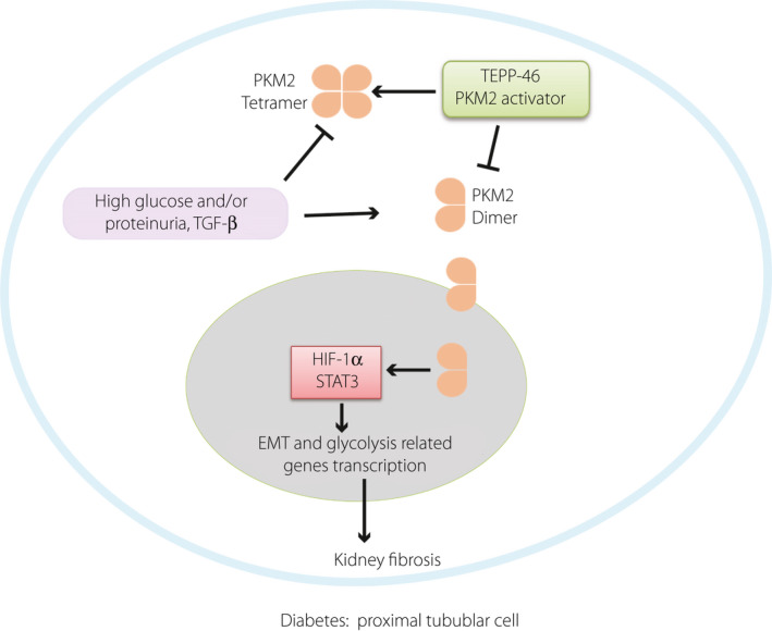
AIMS/INTRODUCTION: Tubulointerstitial fibrosis is a hallmark of diabetic nephropathy and is associated with an epithelial-to-mesenchymal transition (EMT) program and aberrant glycolysis. Dimeric pyruvate kinase (PK) M2 (PKM2) acts as a key protein kinase in aberrant glycolysis by promoting the accumulation of hypoxia-inducible factor (HIF)-1α, while tetrameric PKM2 functions as a pyruvate kinase in oxidative phosphorylation. The aim of the research is to study the effect of PKM2 tetramer activation on preventing kidney fibrosis via suppression of aberrant glycolysis and the EMT program.
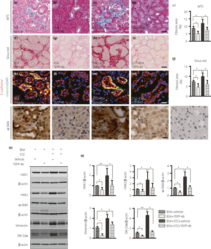
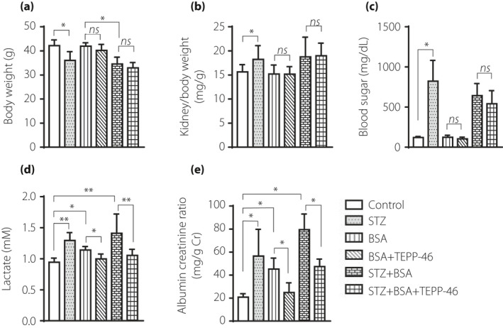
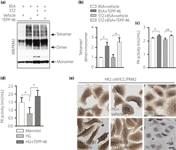
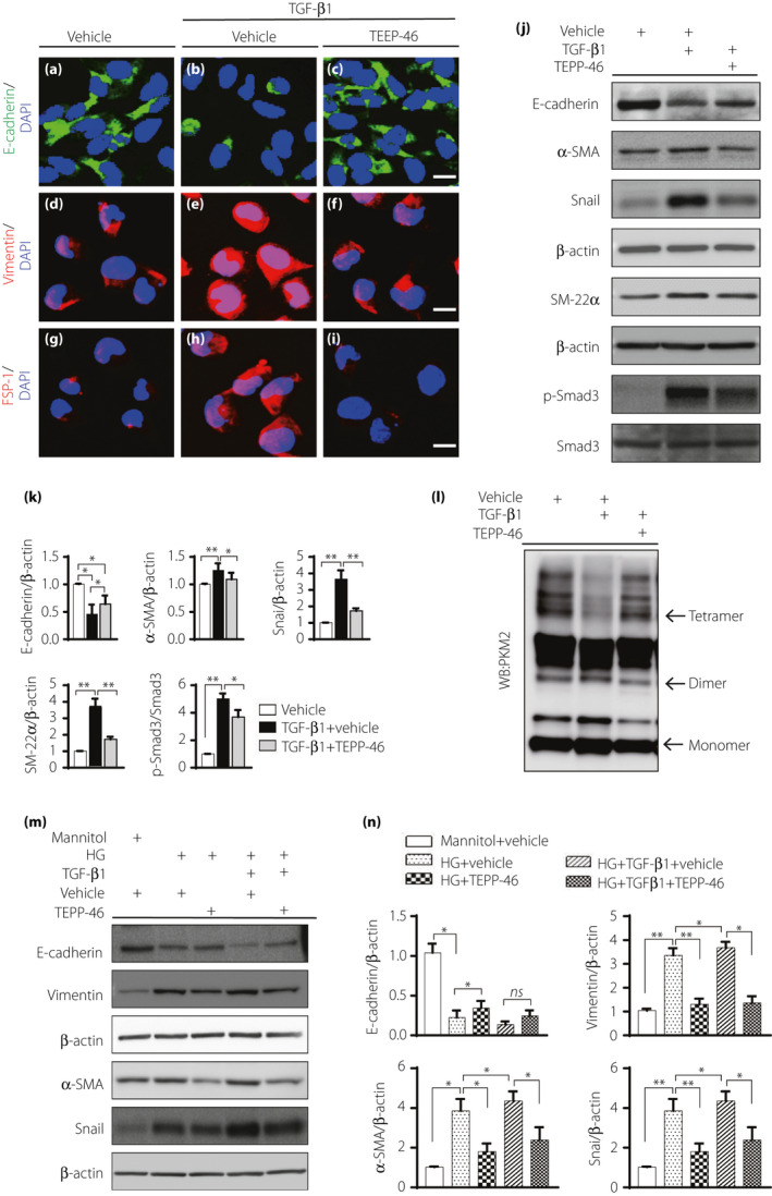
In vivo: Streptozotocin (STZ) was utilized to induce diabetes in 8-week-old CD-1 mice; 4 weeks after diabetes induction, proteinuria-induced kidney fibrosis was developed by intraperitoneal injection of bovine serum albumin (BSA: 0.3 g/30 g BW) for 14 days; The PKM2 activator TEPP-46 was also administered orally simultaneously. In vitro: HK2 cells were co-treated with high-glucose media or/and TGF-β1 and TEPP46 for 48 h, cellular protein was extracted for evaluation.
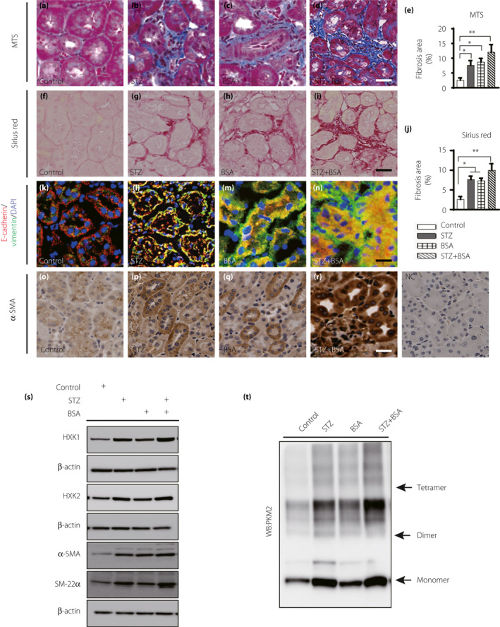
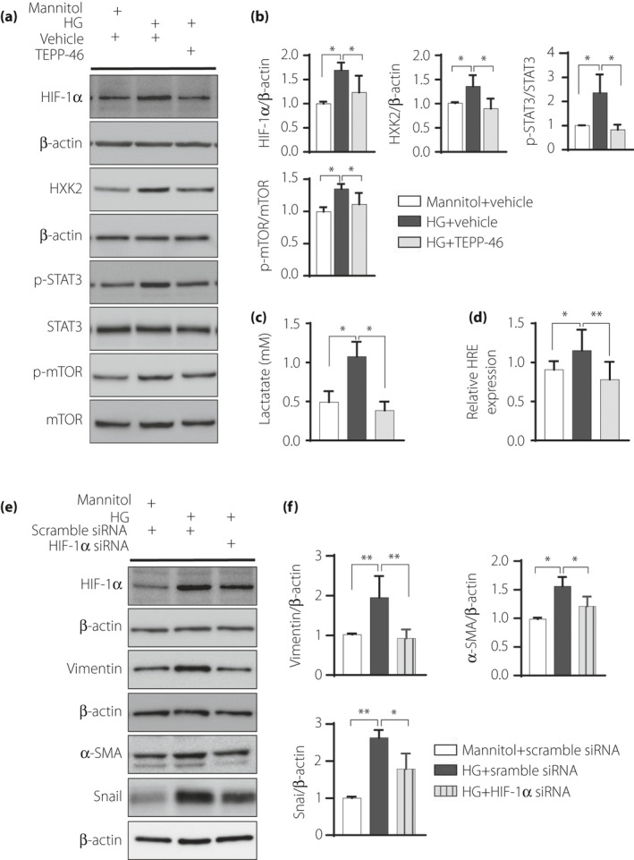
Diabetic mice developed kidney fibrosis associated with aberrant glycolysis and EMT; BSA injection accelerated kidney fibrosis in both the control and diabetic mice; TEPP-46 rescued the kidney fibrosis. In HK2 cells, TEPP-46 suppressed the EMT program induced by TGF-β1 and/or high-glucose incubation. TEPP-46-induced PKM2 tetramer formation and PK activity resulted in suppression of HIF-1α and lactate accumulation. Specific siRNA-mediated knockdown of HIF-1α expression diminished high glucose-induced mesenchymal protein levels.
PKM2 activation could restore the tubular phenotype via suppression of the EMT program and aberrant glycolysis, providing an alternative target to mitigate fibrosis in diabetic kidneys.
目的/引言:肾小管间质纤维化是糖尿病肾病的标志,与上皮间质转化(EMT)程序和异常糖酵解有关。二聚丙酮酸激酶(PK)M2(PKM2)通过促进缺氧诱导因子(HIF)-1α的积累,作为异常糖酵解的关键蛋白激酶发挥作用,而四聚体 PKM2 在氧化磷酸化中作为丙酮酸激酶发挥作用。本研究旨在通过抑制异常糖酵解和 EMT 程序来研究激活 PKM2 四聚体对预防肾脏纤维化的作用。
体内:链脲佐菌素(STZ)用于诱导 8 周龄 CD-1 小鼠的糖尿病;糖尿病诱导 4 周后,通过腹腔注射牛血清白蛋白(BSA:0.3g/30g BW)诱导蛋白尿诱导的肾脏纤维化 14 天;同时口服给予 PKM2 激活剂 TEPP-46。体外:HK2 细胞与高糖培养基或/和 TGF-β1 和 TEPP46 共处理 48h,提取细胞蛋白进行评估。
糖尿病小鼠发生与异常糖酵解和 EMT 相关的肾脏纤维化;BSA 注射加速了对照组和糖尿病小鼠的肾脏纤维化;TEPP-46 挽救了肾脏纤维化。在 HK2 细胞中,TEPP-46 抑制了 TGF-β1 和/或高糖孵育诱导的 EMT 程序。TEPP-46 诱导的 PKM2 四聚体形成和 PK 活性导致 HIF-1α 和乳酸积累的抑制。特异性 siRNA 介导的 HIF-1α 表达敲低减少了高葡萄糖诱导的间充质蛋白水平。
PKM2 激活可通过抑制 EMT 程序和异常糖酵解恢复肾小管表型,为减轻糖尿病肾脏纤维化提供了另一种靶标。