Laboratoire de Cartographie fonctionnelle du Cerveau (LCFC), UNI-ULB Neuroscience Institute, Université libre de Bruxelles (ULB), Brussels, Belgium.
Neuropsychology and Functional Neuroimaging Research Unit (UR2NF), Center for Research in Cognition and Neurosciences (CRCN), UNI-ULB Neuroscience Institute, Université libre de Bruxelles (ULB), Brussels, Belgium.
Sci Rep. 2020 Dec 15;10(1):21990. doi: 10.1038/s41598-020-76201-3.
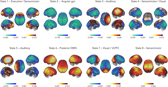
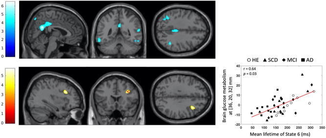
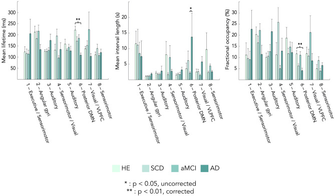
Human brain activity is intrinsically organized into resting-state networks (RSNs) that transiently activate or deactivate at the sub-second timescale. Few neuroimaging studies have addressed how Alzheimer's disease (AD) affects these fast temporal brain dynamics, and how they relate to the cognitive, structural and metabolic abnormalities characterizing AD. We aimed at closing this gap by investigating both brain structure and function using magnetoencephalography (MEG) and hybrid positron emission tomography-magnetic resonance (PET/MR) in 10 healthy elders, 10 patients with subjective cognitive decline (SCD), 10 patients with amnestic mild cognitive impairment (aMCI) and 10 patients with typical Alzheimer's disease with dementia (AD). The fast activation/deactivation state dynamics of RSNs were assessed using hidden Markov modeling (HMM) of power envelope fluctuations at rest measured with MEG. Correlations were sought between temporal properties of HMM states and participants' cognitive test scores, whole hippocampal grey matter volume and regional brain glucose metabolism. The posterior default-mode network (DMN) was less often activated and for shorter durations in AD patients than matched healthy elders. No significant difference was found in patients with SCD or aMCI. The time spent by participants in the activated posterior DMN state did not correlate significantly with cognitive scores, nor with the whole hippocampal volume. However, it correlated positively with the regional glucose consumption in the right dorsolateral prefrontal cortex (DLPFC). AD patients present alterations of posterior DMN power activation dynamics at rest that identify an additional electrophysiological correlate of AD-related synaptic and neural dysfunction. The right DLPFC may play a causal role in the activation of the posterior DMN, possibly linked to the occurrence of mind wandering episodes. As such, these data might suggest a neural correlate of the decrease in mind wandering episodes reported in pathological aging.
人脑活动在亚秒时间尺度上固有地组织为静息状态网络 (RSN),这些网络会短暂激活或失活。很少有神经影像学研究探讨阿尔茨海默病 (AD) 如何影响这些快速的大脑动态,以及它们与 AD 特征的认知、结构和代谢异常有何关系。我们旨在通过使用脑磁图 (MEG) 和正电子发射断层扫描-磁共振 (PET/MR) 联合研究 10 名健康老年人、10 名有主观认知减退 (SCD) 的患者、10 名有遗忘型轻度认知障碍 (aMCI) 的患者和 10 名有典型痴呆症的阿尔茨海默病患者,来填补这一空白。使用 MEG 测量的静息状态下功率包络波动的隐马尔可夫模型 (HMM) 评估了 RSN 的快速激活/失活状态动态。在参与者的认知测试分数、整个海马灰质体积和区域脑葡萄糖代谢之间寻找 HMM 状态的时间特性之间的相关性。与匹配的健康老年人相比,AD 患者的后默认模式网络 (DMN) 较少被激活,且持续时间较短。在 SCD 或 aMCI 患者中未发现显著差异。参与者处于激活的后 DMN 状态的时间与认知评分、整个海马体积均无显著相关性。然而,它与右侧背外侧前额叶皮层 (DLPFC) 的区域葡萄糖消耗呈正相关。AD 患者在静息时出现后 DMN 功率激活动力学改变,这确定了与 AD 相关的突触和神经功能障碍的另一个电生理相关性。右侧 DLPFC 可能在后 DMN 的激活中发挥因果作用,可能与思维漫游事件的发生有关。因此,这些数据可能表明病理性衰老中报告的思维漫游事件减少的神经相关性。