Zhang Jinghang, Pei Lihong, Zang Dan, Xue Yun, Wang Xiaohui, Chen Yiyang, Li Jinsong, Yu Jian, Gao Qingzu, Di Wenyu, Cui Chaochu, Su Wei, Wang Xianwei
Department of Pathology, The First Affiliated Hospital of Xinxiang Medical University, Xinxiang, China.
Key Laboratory of Lymphohematopoietic Tumor in Xinxiang, Xinxiang Medical University, Xinxiang, China.
Front Aging Neurosci. 2020 Nov 19;12:512097. doi: 10.3389/fnagi.2020.512097. eCollection 2020.
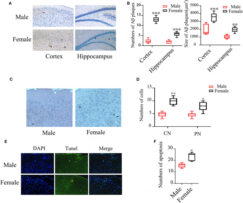
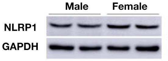
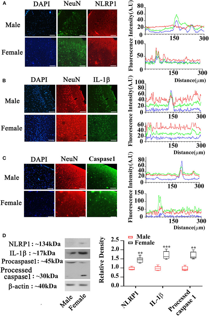
There is a significant gender difference in the incidence and symptoms of Alzheimer's disease (AD), but its mechanisms are not completely understood. Recent studies showed that NLRP1 inflammasome was overexpressed in females under some pathological conditions such as nodular melanoma. Whether NLRP1 signals have a gender difference in AD has not been elucidated. This study was designed to investigate gender difference on the expressions of NLRP1 signals including NLRP1, Capase-1 and IL-1β in the brains of APP/PS1 mice. Female and male APP/PS1 mice (30-weeks-old) were used in this study. Amyloid-β (Aβ) plaques were stained with Congo red dye and cell apoptosis was detected by TUNEL staining. Expressions of NLRP1, Capase-1 and IL-1β were measured by immunofluorescent staining and Western blotting assay. The numbers of Aβ plaques in cortex and hippocampus and neuronal apoptosis in cortex were 4 and 2-folds in females than males, respectively ( < 0.001). The average size of Aβ plaques in both cortex (females: 3527.11 ± 539.88 μm vs. males: 1920.44 ± 638.49 μm) and hippocampus (females: 1931 ± 308.61 μm vs. males: 1038.55 ± 220.40 μm) were also larger in females than males ( < 0.01). More interestingly, expressions of NLRP1, Caspase-1, and IL-1β were markedly increased in the cortex of females as compared with males. These findings show that NLRP1 signals are higher in brains of female APP/PS1 mice than males, which may be related to the gender differences of AD.
阿尔茨海默病(AD)的发病率和症状存在显著的性别差异,但其机制尚未完全明确。最近的研究表明,在某些病理条件下,如结节性黑色素瘤,NLRP1炎性小体在女性中过度表达。NLRP1信号在AD中是否存在性别差异尚未阐明。本研究旨在探讨APP/PS1小鼠大脑中NLRP1信号(包括NLRP1、半胱天冬酶-1和白细胞介素-1β)表达的性别差异。本研究使用了30周龄的雌性和雄性APP/PS1小鼠。用刚果红染料对淀粉样β蛋白(Aβ)斑块进行染色,并通过TUNEL染色检测细胞凋亡。通过免疫荧光染色和蛋白质免疫印迹法检测NLRP1、半胱天冬酶-1和白细胞介素-1β的表达。雌性小鼠皮质和海马中的Aβ斑块数量以及皮质中的神经元凋亡数量分别是雄性小鼠的4倍和2倍(P<0.001)。雌性小鼠皮质(雌性:3527.11±539.88μm,雄性:1920.44±638.49μm)和海马(雌性:1931±308.61μm,雄性:1038.55±220.40μm)中Aβ斑块的平均大小也大于雄性小鼠(P<0.01)。更有趣的是,与雄性小鼠相比,雌性小鼠皮质中NLRP1、半胱天冬酶-1和白细胞介素-1β的表达明显增加。这些发现表明,雌性APP/PS1小鼠大脑中的NLRP1信号高于雄性小鼠,这可能与AD的性别差异有关。