• 文献检索
  • 文档翻译
  • 深度研究
  • 学术资讯
  • Suppr Zotero 插件Zotero 插件
  • 邀请有礼
  • 套餐&价格
  • 历史记录
应用&插件
Suppr Zotero 插件Zotero 插件浏览器插件Mac 客户端Windows 客户端微信小程序
定价
高级版会员购买积分包购买API积分包
服务
文献检索文档翻译深度研究API 文档MCP 服务
关于我们
关于 Suppr公司介绍联系我们用户协议隐私条款
关注我们

Suppr 超能文献

核心技术专利:CN118964589B侵权必究
粤ICP备2023148730 号-1Suppr @ 2026

文献检索

告别复杂PubMed语法,用中文像聊天一样搜索,搜遍4000万医学文献。AI智能推荐,让科研检索更轻松。

立即免费搜索

文件翻译

保留排版,准确专业,支持PDF/Word/PPT等文件格式,支持 12+语言互译。

免费翻译文档

深度研究

AI帮你快速写综述,25分钟生成高质量综述,智能提取关键信息,辅助科研写作。

立即免费体验

以全层组织病理学信息为参考的MRI相关性研究中定义的原发性前列腺癌患者前列腺内大体肿瘤体积的各向同性扩展。

Isotropic Expansion of the Intraprostatic Gross Tumor Volume of Primary Prostate Cancer Patients Defined in MRI-A Correlation Study With Whole Mount Histopathological Information as Reference.

作者信息

Kramer Maria, Spohn Simon K B, Kiefer Selina, Ceci Lara, Sigle August, Oerther Benedict, Schultze-Seemann Wolfgang, Gratzke Christian, Bock Michael, Bamberg Fabian, Grosu Anca L, Benndorf Matthias, Zamboglou Constantinos

机构信息

Department of Radiation Oncology, Medical Center-University of Freiburg, Faculty of Medicine, University of Freiburg, Freiburg, Germany.

Institute of Surgical Pathology, Medical Center-University of Freiburg, Faculty of Medicine, University of Freiburg, Freiburg, Germany.

出版信息

Front Oncol. 2020 Nov 23;10:596756. doi: 10.3389/fonc.2020.596756. eCollection 2020.

DOI:10.3389/fonc.2020.596756
PMID:33330088
原文链接:https://pmc.ncbi.nlm.nih.gov/articles/PMC7719800/
Abstract

INTRODUCTION

An accurate delineation of the intraprostatic gross tumor volume (GTV) is of importance for focal treatment in patients with primary prostate cancer (PCa). Multiparametric MRI (mpMRI) is the standard of care for lesion detection but has been shown to underestimate GTV. This study investigated how far the GTV has to be expanded in MRI in order to reach concordance with the histopathological reference and whether this strategy is practicable in clinical routine.

PATIENTS AND METHODS

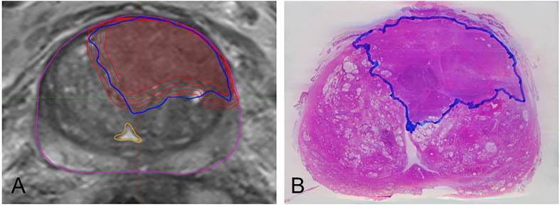
Twenty-two patients with planned prostatectomy and preceded 3 Tesla mpMRI were prospectively examined. After surgery, PCa contours delineated on histopathological slides (GTV-Histo) were superimposed on MRI using imaging as support for co-registration. According to the PI-RADSv2 classification, GTV was manually delineated in MRI (GTV-MRI) by two experts in consensus. For volumetric analysis, we compared GTV-MRI and GTV-Histo. Subsequently, we isotropically enlarged GTV-MRI in 1 mm increments within the prostate and also compared those with GTV-Histo regarding the absolute volumes. For evaluating the spatial accuracy, we considered the coverage ratio of GTV-Histo, the Sørensen-Dice coefficient (DSC), as well as the contact with the urethra.

RESULTS

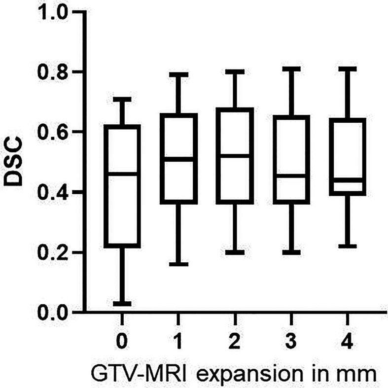
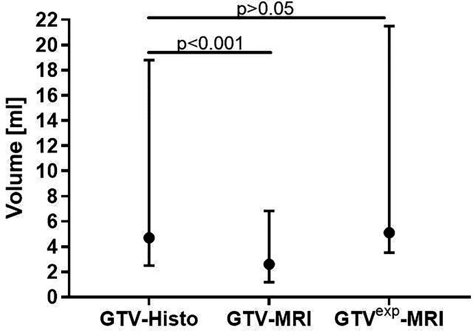
In 19 of 22 patients MRI underestimated the intraprostatic tumor volume compared to histopathological reference: median GTV-Histo (4.7 cm, IQR: 2.5-18.8) was significantly (p<0.001) lager than median GTV-MRI (2.6 cm, IQR: 1.2-6.9). A median expansion of 1 mm (range: 0-4 mm) adjusted the initial GTV-MRI to at least the volume of GTV-Histo (GTV-MRI). Original GTV-MRI and expansion with 1, 2, 3, and 4 mm covered in median 39% (IQR: 2%-78%), 62% (10%-91%), 70% (15%-95%), 80% (21-100), 87% (25%-100%) of GTV-Histo, respectively. Best DSC (median: 0.54) between GTV-Histo and GTV-MRI was achieved by median expansion of 2 mm. The urethra was covered by initial GTVs-MRI in eight patients (36%). After applying an expansion with 2 mm the urethra was covered in one more patient by GTV-MRI.

CONCLUSION

Using histopathology as reference, we demonstrated that MRI underestimates intraprostatic tumor volume. A 2 mm-expansion may improve accurate GTV-delineation while respecting the balance between histological tumor coverage and overtreatment.

摘要

引言

准确描绘前列腺内大体肿瘤体积(GTV)对于原发性前列腺癌(PCa)患者的局部治疗至关重要。多参数MRI(mpMRI)是病变检测的标准护理方法,但已被证明会低估GTV。本研究调查了在MRI中GTV需要扩大多少才能与组织病理学参考值一致,以及这种策略在临床常规中是否可行。

患者和方法

前瞻性检查了22例计划进行前列腺切除术且术前行3特斯拉mpMRI检查的患者。手术后,将组织病理学切片上勾勒出的PCa轮廓(GTV-Histo)叠加在MRI上,并使用成像作为共配准的支持。根据PI-RADSv2分类,由两位专家达成共识,在MRI中手动勾勒出GTV(GTV-MRI)。对于体积分析,我们比较了GTV-MRI和GTV-Histo。随后,我们在前列腺内以1毫米的增量各向同性地扩大GTV-MRI,并将其与GTV-Histo的绝对体积进行比较。为了评估空间准确性,我们考虑了GTV-Histo的覆盖率、Sørensen-Dice系数(DSC)以及与尿道的接触情况。

结果

与组织病理学参考值相比,22例患者中有19例MRI低估了前列腺内肿瘤体积:GTV-Histo的中位数(4.7厘米,四分位间距:2.5-18.8)显著大于(p<0.001)GTV-MRI的中位数(2.6厘米,四分位间距:1.2-6.9)。中位数扩大1毫米(范围:0-4毫米)可将初始GTV-MRI至少调整到GTV-Histo(GTV-MRI)的体积。原始GTV-MRI以及扩大1、2、3和4毫米后的GTV分别在中位数上覆盖了GTV-Histo的39%(四分位间距:2%-78%)、62%(10%-91%)、70%(15%-95%)、80%(21%-100%)、87%(25%-100%)。GTV-Histo与GTV-MRI之间最佳的DSC(中位数:0.54)是通过中位数扩大2毫米实现的。8例患者(36%)的初始GTV-MRI覆盖了尿道。在应用2毫米的扩大后,GTV-MRI又多覆盖了1例患者的尿道。

结论

以组织病理学为参考,我们证明了MRI低估了前列腺内肿瘤体积。2毫米的扩大可能会在尊重组织学肿瘤覆盖和过度治疗之间的平衡的同时,改善GTV的准确勾勒。

https://cdn.ncbi.nlm.nih.gov/pmc/blobs/0376/7719800/b70554d929aa/fonc-10-596756-g003.jpg
https://cdn.ncbi.nlm.nih.gov/pmc/blobs/0376/7719800/bb8e68374329/fonc-10-596756-g001.jpg
https://cdn.ncbi.nlm.nih.gov/pmc/blobs/0376/7719800/69e9b7dbb5d7/fonc-10-596756-g002.jpg
https://cdn.ncbi.nlm.nih.gov/pmc/blobs/0376/7719800/b70554d929aa/fonc-10-596756-g003.jpg
https://cdn.ncbi.nlm.nih.gov/pmc/blobs/0376/7719800/bb8e68374329/fonc-10-596756-g001.jpg
https://cdn.ncbi.nlm.nih.gov/pmc/blobs/0376/7719800/69e9b7dbb5d7/fonc-10-596756-g002.jpg
https://cdn.ncbi.nlm.nih.gov/pmc/blobs/0376/7719800/b70554d929aa/fonc-10-596756-g003.jpg

相似文献

1
Isotropic Expansion of the Intraprostatic Gross Tumor Volume of Primary Prostate Cancer Patients Defined in MRI-A Correlation Study With Whole Mount Histopathological Information as Reference.以全层组织病理学信息为参考的MRI相关性研究中定义的原发性前列腺癌患者前列腺内大体肿瘤体积的各向同性扩展。
Front Oncol. 2020 Nov 23;10:596756. doi: 10.3389/fonc.2020.596756. eCollection 2020.
2
Comparison of Ga-HBED-CC PSMA-PET/CT and multiparametric MRI for gross tumour volume detection in patients with primary prostate cancer based on slice by slice comparison with histopathology.基于与组织病理学逐片比较,比较镓-羟基苄基乙二胺-N,N'-二乙酸-环糊精前列腺特异性膜抗原正电子发射断层扫描/计算机断层扫描(Ga-HBED-CC PSMA-PET/CT)和多参数磁共振成像(MRI)在原发性前列腺癌患者中检测大体肿瘤体积的情况。
Theranostics. 2017 Jan 1;7(1):228-237. doi: 10.7150/thno.16638. eCollection 2017.
3
[Ga-]PSMA-11 PET/CT and multiparametric MRI for gross tumor volume delineation in a slice by slice analysis with whole mount histopathology as a reference standard - Implications for focal radiotherapy planning in primary prostate cancer.[镓-68]PSMA-11 PET/CT 和多参数 MRI 用于逐层分析勾画大体肿瘤靶区,以全器官病理为参考标准 - 对原发性前列腺癌调强放疗计划的影响。
Radiother Oncol. 2019 Dec;141:214-219. doi: 10.1016/j.radonc.2019.07.005. Epub 2019 Aug 17.
4
Influence of Urethra Sparing on Tumor Control Probability and Normal Tissue Complication Probability in Focal Dose Escalated Hypofractionated Radiotherapy: A Planning Study Based on Histopathology Reference.保留尿道对局部剂量递增的低分割放疗中肿瘤控制概率和正常组织并发症概率的影响:一项基于组织病理学参考的计划研究
Front Oncol. 2021 May 14;11:652678. doi: 10.3389/fonc.2021.652678. eCollection 2021.
5
Comparison of Manual and Semi-Automatic [F]PSMA-1007 PET Based Contouring Techniques for Intraprostatic Tumor Delineation in Patients With Primary Prostate Cancer and Validation With Histopathology as Standard of Reference.基于手动和半自动[F]PSMA - 1007 PET的轮廓描绘技术在原发性前列腺癌患者前列腺内肿瘤勾画中的比较以及以组织病理学为参考标准的验证
Front Oncol. 2020 Dec 7;10:600690. doi: 10.3389/fonc.2020.600690. eCollection 2020.
6
Focal dose escalation for prostate cancer using Ga-HBED-CC PSMA PET/CT and MRI: a planning study based on histology reference.基于组织学参考的 Ga-HBED-CC PSMA PET/CT 和 MRI 引导前列腺癌焦点剂量递增:一项计划研究。
Radiat Oncol. 2018 May 2;13(1):81. doi: 10.1186/s13014-018-1036-8.
7
Intraindividual comparison between Ga-PSMA-PET/CT and mpMRI for intraprostatic tumor delineation in patients with primary prostate cancer: a retrospective analysis in 101 patients.在原发性前列腺癌患者中,Ga-PSMA-PET/CT 与 mpMRI 对前列腺内肿瘤勾画的个体内比较:101 例患者的回顾性分析。
Eur J Nucl Med Mol Imaging. 2020 Nov;47(12):2796-2803. doi: 10.1007/s00259-020-04827-6. Epub 2020 Apr 28.
8
Intraindividual Comparison Between [F] PSMA-1007 PET/CT and Multiparametric MRI for Radiotherapy Planning in Primary Prostate Cancer Patients.[F] PSMA - 1007 PET/CT与多参数MRI在原发性前列腺癌患者放疗计划中的个体内比较
Front Oncol. 2022 Jul 14;12:880042. doi: 10.3389/fonc.2022.880042. eCollection 2022.
9
Radiomic features from PSMA PET for non-invasive intraprostatic tumor discrimination and characterization in patients with intermediate- and high-risk prostate cancer - a comparison study with histology reference.基于 PSMA PET 的放射组学特征在中高危前列腺癌患者中进行前列腺内肿瘤的非侵入性鉴别和特征分析——与组织学参考的比较研究。
Theranostics. 2019 Apr 13;9(9):2595-2605. doi: 10.7150/thno.32376. eCollection 2019.
10
MRI versus ⁶⁸Ga-PSMA PET/CT for gross tumour volume delineation in radiation treatment planning of primary prostate cancer.在原发性前列腺癌放射治疗计划中,MRI与⁶⁸Ga-PSMA PET/CT在大体肿瘤体积勾画方面的比较
Eur J Nucl Med Mol Imaging. 2016 May;43(5):889-897. doi: 10.1007/s00259-015-3257-5. Epub 2015 Nov 23.

引用本文的文献

1
Histopathology-validated gross tumor volume delineations of intraprostatic lesions using PSMA-positron emission tomography/multiparametric magnetic resonance imaging.使用前列腺特异性膜抗原正电子发射断层扫描/多参数磁共振成像对前列腺内病变进行组织病理学验证的大体肿瘤体积勾画
Phys Imaging Radiat Oncol. 2024 Aug 22;31:100633. doi: 10.1016/j.phro.2024.100633. eCollection 2024 Jul.
2
Target Volume Optimization for Localized Prostate Cancer.局限性前列腺癌的靶区优化。
Pract Radiat Oncol. 2024 Nov-Dec;14(6):522-540. doi: 10.1016/j.prro.2024.06.006. Epub 2024 Jul 15.
3
Advantage of whole-mount histopathology in prostate cancer: current applications and future prospects.

本文引用的文献

1
Intraindividual comparison between Ga-PSMA-PET/CT and mpMRI for intraprostatic tumor delineation in patients with primary prostate cancer: a retrospective analysis in 101 patients.在原发性前列腺癌患者中,Ga-PSMA-PET/CT 与 mpMRI 对前列腺内肿瘤勾画的个体内比较:101 例患者的回顾性分析。
Eur J Nucl Med Mol Imaging. 2020 Nov;47(12):2796-2803. doi: 10.1007/s00259-020-04827-6. Epub 2020 Apr 28.
2
Validation of different PSMA-PET/CT-based contouring techniques for intraprostatic tumor definition using histopathology as standard of reference.使用组织病理学作为参考标准,验证不同基于 PSMA-PET/CT 的前列腺内肿瘤勾画技术。
Radiother Oncol. 2019 Dec;141:208-213. doi: 10.1016/j.radonc.2019.07.002. Epub 2019 Aug 17.
3
前列腺癌全器官病理检查的优势:当前的应用与未来的前景。
BMC Cancer. 2024 Apr 11;24(1):448. doi: 10.1186/s12885-024-12071-6.
4
Intraindividual Comparison Between [F] PSMA-1007 PET/CT and Multiparametric MRI for Radiotherapy Planning in Primary Prostate Cancer Patients.[F] PSMA - 1007 PET/CT与多参数MRI在原发性前列腺癌患者放疗计划中的个体内比较
Front Oncol. 2022 Jul 14;12:880042. doi: 10.3389/fonc.2022.880042. eCollection 2022.
5
Explainable AI for CNN-based prostate tumor segmentation in multi-parametric MRI correlated to whole mount histopathology.基于卷积神经网络的多参数 MRI 前列腺肿瘤分割的可解释人工智能与全组织病理相关。
Radiat Oncol. 2022 Apr 2;17(1):65. doi: 10.1186/s13014-022-02035-0.
6
PSMA-PET/MRI-Based Focal Dose Escalation in Patients with Primary Prostate Cancer Treated with Stereotactic Body Radiation Therapy (HypoFocal-SBRT): Study Protocol of a Randomized, Multicentric Phase III Trial.基于PSMA-PET/MRI的立体定向体部放射治疗(HypoFocal-SBRT)原发性前列腺癌患者的局部剂量递增:一项随机、多中心III期试验的研究方案
Cancers (Basel). 2021 Nov 18;13(22):5795. doi: 10.3390/cancers13225795.
7
Tumour growth rates of prostate cancer during active surveillance: is there a difference between MRI-visible low and intermediate-risk disease?主动监测期间前列腺癌的肿瘤生长速度:MRI 可见的低危和中危疾病之间是否存在差异?
Br J Radiol. 2022 Mar 1;95(1131):20210321. doi: 10.1259/bjr.20210321. Epub 2021 Jul 8.
[Ga-]PSMA-11 PET/CT and multiparametric MRI for gross tumor volume delineation in a slice by slice analysis with whole mount histopathology as a reference standard - Implications for focal radiotherapy planning in primary prostate cancer.
[镓-68]PSMA-11 PET/CT 和多参数 MRI 用于逐层分析勾画大体肿瘤靶区,以全器官病理为参考标准 - 对原发性前列腺癌调强放疗计划的影响。
Radiother Oncol. 2019 Dec;141:214-219. doi: 10.1016/j.radonc.2019.07.005. Epub 2019 Aug 17.
4
Detection of Individual Prostate Cancer Foci via Multiparametric Magnetic Resonance Imaging.通过多参数磁共振成像检测个体前列腺癌病灶。
Eur Urol. 2019 May;75(5):712-720. doi: 10.1016/j.eururo.2018.11.031. Epub 2018 Dec 1.
5
Focal dose escalation for prostate cancer using Ga-HBED-CC PSMA PET/CT and MRI: a planning study based on histology reference.基于组织学参考的 Ga-HBED-CC PSMA PET/CT 和 MRI 引导前列腺癌焦点剂量递增:一项计划研究。
Radiat Oncol. 2018 May 2;13(1):81. doi: 10.1186/s13014-018-1036-8.
6
The dose distribution in dominant intraprostatic tumour lesions defined by multiparametric MRI and PSMA PET/CT correlates with the outcome in patients treated with primary radiation therapy for prostate cancer.由多参数MRI和PSMA PET/CT定义的前列腺内主要肿瘤病灶中的剂量分布与接受前列腺癌原发性放射治疗患者的预后相关。
Radiat Oncol. 2018 Apr 12;13(1):65. doi: 10.1186/s13014-018-1014-1.
7
MRI-Targeted or Standard Biopsy for Prostate-Cancer Diagnosis.MRI 靶向或标准活检用于前列腺癌诊断。
N Engl J Med. 2018 May 10;378(19):1767-1777. doi: 10.1056/NEJMoa1801993. Epub 2018 Mar 18.
8
Effect of Standard vs Dose-Escalated Radiation Therapy for Patients With Intermediate-Risk Prostate Cancer: The NRG Oncology RTOG 0126 Randomized Clinical Trial.标准剂量与剂量递增放疗治疗中危前列腺癌患者的效果:NRG 肿瘤学 RTOG 0126 随机临床试验。
JAMA Oncol. 2018 Jun 14;4(6):e180039. doi: 10.1001/jamaoncol.2018.0039.
9
Standard whole prostate gland radiotherapy with and without lesion boost in prostate cancer: Toxicity in the FLAME randomized controlled trial.标准全前列腺放疗联合或不联合病灶加量放疗治疗前列腺癌:FLAME 随机对照试验中的毒性反应。
Radiother Oncol. 2018 Apr;127(1):74-80. doi: 10.1016/j.radonc.2017.12.022. Epub 2018 Jan 11.
10
Androgen Suppression Combined with Elective Nodal and Dose Escalated Radiation Therapy (the ASCENDE-RT Trial): An Analysis of Survival Endpoints for a Randomized Trial Comparing a Low-Dose-Rate Brachytherapy Boost to a Dose-Escalated External Beam Boost for High- and Intermediate-risk Prostate Cancer.雄激素抑制联合选择性淋巴结及剂量递增放射治疗(ASCENDE-RT试验):一项针对高风险和中风险前列腺癌的随机试验的生存终点分析,该试验比较了低剂量率近距离放疗增敏与剂量递增外照射增敏。
Int J Radiat Oncol Biol Phys. 2017 Jun 1;98(2):275-285. doi: 10.1016/j.ijrobp.2016.11.026. Epub 2016 Nov 24.