• 文献检索
  • 文档翻译
  • 深度研究
  • 学术资讯
  • Suppr Zotero 插件Zotero 插件
  • 邀请有礼
  • 套餐&价格
  • 历史记录
应用&插件
Suppr Zotero 插件Zotero 插件浏览器插件Mac 客户端Windows 客户端微信小程序
定价
高级版会员购买积分包购买API积分包
服务
文献检索文档翻译深度研究API 文档MCP 服务
关于我们
关于 Suppr公司介绍联系我们用户协议隐私条款
关注我们

Suppr 超能文献

核心技术专利:CN118964589B侵权必究
粤ICP备2023148730 号-1Suppr @ 2026

文献检索

告别复杂PubMed语法,用中文像聊天一样搜索,搜遍4000万医学文献。AI智能推荐,让科研检索更轻松。

立即免费搜索

文件翻译

保留排版,准确专业,支持PDF/Word/PPT等文件格式,支持 12+语言互译。

免费翻译文档

深度研究

AI帮你快速写综述,25分钟生成高质量综述,智能提取关键信息,辅助科研写作。

立即免费体验

基于手动和半自动[F]PSMA - 1007 PET的轮廓描绘技术在原发性前列腺癌患者前列腺内肿瘤勾画中的比较以及以组织病理学为参考标准的验证

Comparison of Manual and Semi-Automatic [F]PSMA-1007 PET Based Contouring Techniques for Intraprostatic Tumor Delineation in Patients With Primary Prostate Cancer and Validation With Histopathology as Standard of Reference.

作者信息

Spohn Simon K B, Kramer Maria, Kiefer Selina, Bronsert Peter, Sigle August, Schultze-Seemann Wolfgang, Jilg Cordula A, Sprave Tanja, Ceci Lara, Fassbender Thomas F, Nicolay Nils H, Ruf Juri, Grosu Anca L, Zamboglou Constantinos

机构信息

Department of Radiation Oncology, Medical Center-University of Freiburg, Faculty of Medicine, University of Freiburg, Freiburg, Germany.

German Cancer Consortium (DKTK), Partner Site Freiburg, Freiburg, Germany.

出版信息

Front Oncol. 2020 Dec 7;10:600690. doi: 10.3389/fonc.2020.600690. eCollection 2020.

DOI:10.3389/fonc.2020.600690
PMID:33365271
原文链接:https://pmc.ncbi.nlm.nih.gov/articles/PMC7750498/
Abstract

PURPOSE

Accurate contouring of intraprostatic gross tumor volume (GTV) is pivotal for successful delivery of focal therapies and for biopsy guidance in patients with primary prostate cancer (PCa). Contouring of GTVs, using 18-Fluor labeled tracer prostate specific membrane antigen positron emission tomography ([F]PSMA-1007/PET) has not been examined yet.

PATIENTS AND METHODS

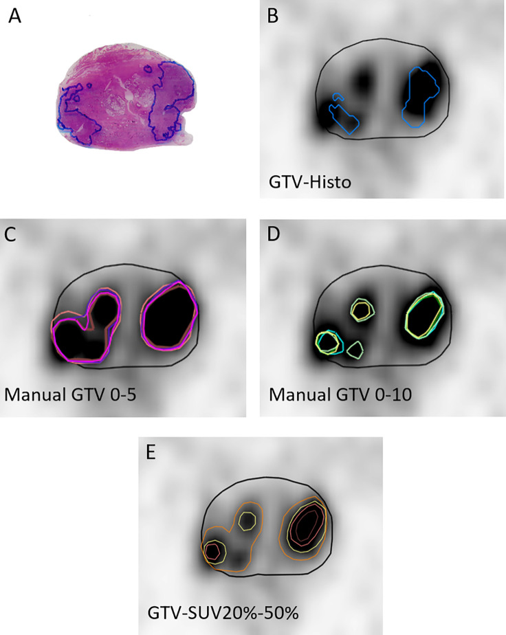
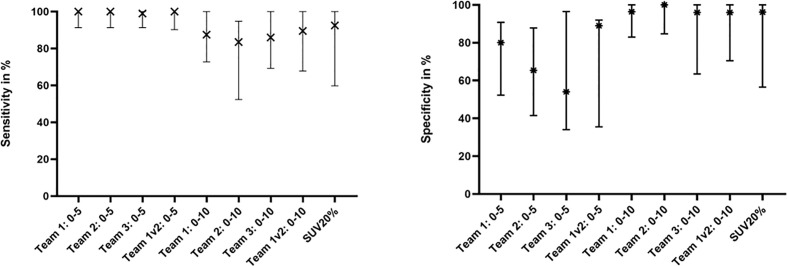
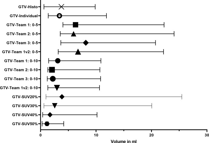
Ten Patients with primary PCa who underwent [F]PSMA-1007 PET followed by radical prostatectomy were prospectively enrolled. Coregistered histopathological gross tumor volume (GTV-Histo) was used as standard of reference. PSMA-PET images were contoured on two ways: (1) manual contouring with PET scaling SUVmin-max: 0-10 was performed by three teams with different levels of experience. Team 1 repeated contouring at a different time point, resulting in n = 4 manual contours. (2) Semi-automatic contouring approaches using SUVmax thresholds of 20-50% were performed. Interobserver agreement was assessed for manual contouring by calculating the Dice Similarity Coefficient (DSC) and for all approaches sensitivity, specificity were calculated by dividing the prostate in each CT slice into four equal quadrants under consideration of histopathology as standard of reference.

RESULTS

Manual contouring yielded an excellent interobserver agreement with a median DSC of 0.90 (range 0.87-0.94). Volumes derived from scaling SUVmin-max 0-10 showed no statistically significant difference from GTV-Histo and high sensitivities (median 87%, range 84-90%) and specificities (median 96%, range 96-100%). GTVs using semi-automatic segmentation applying a threshold of 20-40% of SUVmax showed no significant difference in absolute volumes to GTV-Histo, GTV-SUV50% was significantly smaller. Best performing semi-automatic contour (GTV-SUV20%) achieved high sensitivity (median 93%) and specificity (median 96%). There was no statistically significant difference to SUVmin-max 0-10.

CONCLUSION

Manual contouring with PET scaling SUVmin-max 0-10 and semi-automatic contouring applying a threshold of 20% of SUVmax achieved high sensitivities and very high specificities and are recommended for [F]PSMA-1007 PET based focal therapy approaches. Providing high specificities, semi-automatic approaches applying thresholds of 30-40% of SUVmax are recommend for biopsy guidance.

摘要

目的

准确勾勒前列腺内大体肿瘤体积(GTV)对于成功实施聚焦治疗以及原发性前列腺癌(PCa)患者的活检引导至关重要。尚未对使用18F标记示踪剂前列腺特异性膜抗原正电子发射断层扫描([F]PSMA - 1007/PET)勾勒GTV进行研究。

患者与方法

前瞻性纳入10例接受[F]PSMA - 1007 PET检查后行根治性前列腺切除术的原发性PCa患者。将配准的组织病理学大体肿瘤体积(GTV - Histo)用作参考标准。PSMA - PET图像通过两种方式勾勒:(1)由三个经验水平不同的团队进行PET缩放SUVmin - max:0 - 10的手动勾勒。团队1在不同时间点重复勾勒,得到n = 4个手动轮廓。(2)使用SUVmax阈值为20% - 50%的半自动勾勒方法。通过计算骰子相似系数(DSC)评估手动勾勒的观察者间一致性,对于所有方法,以组织病理学为参考标准,将每个CT切片中的前列腺分为四个相等象限来计算敏感性和特异性。

结果

手动勾勒显示观察者间一致性极佳,DSC中位数为0.90(范围0.87 - 0.94)。缩放SUVmin - max 0 - 10得出的体积与GTV - Histo无统计学显著差异,敏感性高(中位数87%,范围84% - 90%),特异性高(中位数96%,范围96% - 100%)。使用SUVmax阈值为20% - 40%的半自动分割得出的GTV在绝对体积上与GTV - Histo无显著差异,GTV - SUV50%显著更小。表现最佳的半自动轮廓(GTV - SUV20%)实现了高敏感性(中位数93%)和特异性(中位数96%)。与SUVmin - max 0 - 10无统计学显著差异。

结论

PET缩放SUVmin - max 0 - 10的手动勾勒和应用SUVmax 20%阈值的半自动勾勒具有高敏感性和非常高的特异性,推荐用于基于[F]PSMA - 1007 PET的聚焦治疗方法。对于活检引导,推荐应用SUVmax 30% - 40%阈值的半自动方法,其具有高特异性。

https://cdn.ncbi.nlm.nih.gov/pmc/blobs/89ab/7750498/a2059dc4e64d/fonc-10-600690-g003.jpg
https://cdn.ncbi.nlm.nih.gov/pmc/blobs/89ab/7750498/fae88cf3ea67/fonc-10-600690-g001.jpg
https://cdn.ncbi.nlm.nih.gov/pmc/blobs/89ab/7750498/b0d2c7331319/fonc-10-600690-g002.jpg
https://cdn.ncbi.nlm.nih.gov/pmc/blobs/89ab/7750498/a2059dc4e64d/fonc-10-600690-g003.jpg
https://cdn.ncbi.nlm.nih.gov/pmc/blobs/89ab/7750498/fae88cf3ea67/fonc-10-600690-g001.jpg
https://cdn.ncbi.nlm.nih.gov/pmc/blobs/89ab/7750498/b0d2c7331319/fonc-10-600690-g002.jpg
https://cdn.ncbi.nlm.nih.gov/pmc/blobs/89ab/7750498/a2059dc4e64d/fonc-10-600690-g003.jpg

相似文献

1
Comparison of Manual and Semi-Automatic [F]PSMA-1007 PET Based Contouring Techniques for Intraprostatic Tumor Delineation in Patients With Primary Prostate Cancer and Validation With Histopathology as Standard of Reference.基于手动和半自动[F]PSMA - 1007 PET的轮廓描绘技术在原发性前列腺癌患者前列腺内肿瘤勾画中的比较以及以组织病理学为参考标准的验证
Front Oncol. 2020 Dec 7;10:600690. doi: 10.3389/fonc.2020.600690. eCollection 2020.
2
Validation of different PSMA-PET/CT-based contouring techniques for intraprostatic tumor definition using histopathology as standard of reference.使用组织病理学作为参考标准,验证不同基于 PSMA-PET/CT 的前列腺内肿瘤勾画技术。
Radiother Oncol. 2019 Dec;141:208-213. doi: 10.1016/j.radonc.2019.07.002. Epub 2019 Aug 17.
3
Comparison of Ga-HBED-CC PSMA-PET/CT and multiparametric MRI for gross tumour volume detection in patients with primary prostate cancer based on slice by slice comparison with histopathology.基于与组织病理学逐片比较,比较镓-羟基苄基乙二胺-N,N'-二乙酸-环糊精前列腺特异性膜抗原正电子发射断层扫描/计算机断层扫描(Ga-HBED-CC PSMA-PET/CT)和多参数磁共振成像(MRI)在原发性前列腺癌患者中检测大体肿瘤体积的情况。
Theranostics. 2017 Jan 1;7(1):228-237. doi: 10.7150/thno.16638. eCollection 2017.
4
Intraprostatic Tumor Segmentation on PSMA PET Images in Patients with Primary Prostate Cancer with a Convolutional Neural Network.基于卷积神经网络的原发性前列腺癌 PSMA PET 图像前列腺内肿瘤分割。
J Nucl Med. 2021 Jun 1;62(6):823-828. doi: 10.2967/jnumed.120.254623. Epub 2020 Oct 30.
5
[Ga-]PSMA-11 PET/CT and multiparametric MRI for gross tumor volume delineation in a slice by slice analysis with whole mount histopathology as a reference standard - Implications for focal radiotherapy planning in primary prostate cancer.[镓-68]PSMA-11 PET/CT 和多参数 MRI 用于逐层分析勾画大体肿瘤靶区,以全器官病理为参考标准 - 对原发性前列腺癌调强放疗计划的影响。
Radiother Oncol. 2019 Dec;141:214-219. doi: 10.1016/j.radonc.2019.07.005. Epub 2019 Aug 17.
6
Ga-PSMA-11 PET, F-PSMA-1007 PET, and MRI for Gross Tumor Volume Delineation in Primary Prostate Cancer: Intermodality and Intertracer Variability.镓-PSMA-11 PET、F-PSMA-1007 PET 和 MRI 用于原发性前列腺癌大体肿瘤体积勾画:多模态和示踪剂间变异性。
Pract Radiat Oncol. 2021 May-Jun;11(3):202-211. doi: 10.1016/j.prro.2020.11.006.
7
Optimal Ga-PSMA and F-PSMA PET window levelling for gross tumour volume delineation in primary prostate cancer.最佳 Ga-PSMA 和 F-PSMA PET 窗位水平用于原发性前列腺癌大体肿瘤体积勾画。
Eur J Nucl Med Mol Imaging. 2021 Apr;48(4):1211-1218. doi: 10.1007/s00259-020-05059-4. Epub 2020 Oct 6.
8
Comparison of Multiple Segmentation Methods for Volumetric Delineation of Primary Prostate Cancer with Prostate-Specific Membrane Antigen-Targeted F-DCFPyL PET/CT.多分割方法在基于前列腺特异性膜抗原靶向 F-DCFPyL PET/CT 的原发性前列腺癌容积勾画中的比较。
J Nucl Med. 2024 Jan 2;65(1):87-93. doi: 10.2967/jnumed.123.266005.
9
Intraindividual Comparison Between [F] PSMA-1007 PET/CT and Multiparametric MRI for Radiotherapy Planning in Primary Prostate Cancer Patients.[F] PSMA - 1007 PET/CT与多参数MRI在原发性前列腺癌患者放疗计划中的个体内比较
Front Oncol. 2022 Jul 14;12:880042. doi: 10.3389/fonc.2022.880042. eCollection 2022.
10
Intraindividual comparison between Ga-PSMA-PET/CT and mpMRI for intraprostatic tumor delineation in patients with primary prostate cancer: a retrospective analysis in 101 patients.在原发性前列腺癌患者中,Ga-PSMA-PET/CT 与 mpMRI 对前列腺内肿瘤勾画的个体内比较:101 例患者的回顾性分析。
Eur J Nucl Med Mol Imaging. 2020 Nov;47(12):2796-2803. doi: 10.1007/s00259-020-04827-6. Epub 2020 Apr 28.

引用本文的文献

1
Comparative Analysis of Ga-Prostate-Specific Membrane Antigen Positron Emission Tomography/Computed Tomography and Multiparametric Magnetic Resonance Imaging for Gross Tumor Volume Delineation in Radiation Therapy Planning of Prostate Cancer.镓-前列腺特异性膜抗原正电子发射断层扫描/计算机断层扫描与多参数磁共振成像在前列腺癌放射治疗计划中大体肿瘤体积勾画的比较分析
Adv Radiat Oncol. 2025 Jun 9;10(8):101821. doi: 10.1016/j.adro.2025.101821. eCollection 2025 Aug.
2
Interpretable multimodal deep learning model for predicting post-surgical international society of urological pathology grade in primary prostate cancer.用于预测原发性前列腺癌术后国际泌尿病理学会分级的可解释多模态深度学习模型
Eur J Nucl Med Mol Imaging. 2025 Apr 4. doi: 10.1007/s00259-025-07248-5.
3

本文引用的文献

1
Optimal Ga-PSMA and F-PSMA PET window levelling for gross tumour volume delineation in primary prostate cancer.最佳 Ga-PSMA 和 F-PSMA PET 窗位水平用于原发性前列腺癌大体肿瘤体积勾画。
Eur J Nucl Med Mol Imaging. 2021 Apr;48(4):1211-1218. doi: 10.1007/s00259-020-05059-4. Epub 2020 Oct 6.
2
Intraindividual comparison between Ga-PSMA-PET/CT and mpMRI for intraprostatic tumor delineation in patients with primary prostate cancer: a retrospective analysis in 101 patients.在原发性前列腺癌患者中,Ga-PSMA-PET/CT 与 mpMRI 对前列腺内肿瘤勾画的个体内比较:101 例患者的回顾性分析。
Eur J Nucl Med Mol Imaging. 2020 Nov;47(12):2796-2803. doi: 10.1007/s00259-020-04827-6. Epub 2020 Apr 28.
3
Prospective comparison of F-PSMA-1007 PET/CT and MRI with histopathology as the reference standard for intraprostatic tumour detection and T-staging of high-risk prostate cancer.以组织病理学为参考标准,对F-PSMA-1007 PET/CT和MRI在前列腺内肿瘤检测及高危前列腺癌T分期中的应用进行前瞻性比较。
Eur J Nucl Med Mol Imaging. 2025 Mar 31. doi: 10.1007/s00259-025-07208-z.
4
Timing and Patterns of Potentially Salvageable Recurrences Following Stereotactic Body Radiation Therapy for Clinically Localized Prostate Cancer Assessed by Preferential Amino Acid Uptake.通过优先氨基酸摄取评估的立体定向体部放射治疗临床局限性前列腺癌后潜在可挽救复发的时间和模式
Cureus. 2025 Jan 25;17(1):e77964. doi: 10.7759/cureus.77964. eCollection 2025 Jan.
5
Can PSMA PET detect intratumour heterogeneity in histological PSMA expression of primary prostate cancer? Analysis of [Ga]Ga-PSMA-11 and [F]PSMA-1007.前列腺特异性膜抗原(PSMA)正电子发射断层扫描(PET)能否检测出原发性前列腺癌组织学PSMA表达中的肿瘤内异质性?对[镓(Ga)]Ga-PSMA-11和[氟(F)]PSMA-1007的分析
Eur J Nucl Med Mol Imaging. 2025 May;52(6):2023-2033. doi: 10.1007/s00259-025-07078-5. Epub 2025 Jan 17.
6
The impact of multicentric datasets for the automated tumor delineation in primary prostate cancer using convolutional neural networks on F-PSMA-1007 PET.多中心数据集对使用卷积神经网络在F-PSMA-1007 PET上进行原发性前列腺癌自动肿瘤勾画的影响。
Radiat Oncol. 2024 Aug 7;19(1):106. doi: 10.1186/s13014-024-02491-w.
7
Target Volume Optimization for Localized Prostate Cancer.局限性前列腺癌的靶区优化。
Pract Radiat Oncol. 2024 Nov-Dec;14(6):522-540. doi: 10.1016/j.prro.2024.06.006. Epub 2024 Jul 15.
8
Accuracy of gross tumour volume delineation with [68Ga]-PSMA-PET compared to histopathology for high-risk prostate cancer.[68Ga]-PSMA-PET 与组织病理学相比对高危前列腺癌大体肿瘤体积勾画的准确性。
Acta Oncol. 2024 Jun 23;63:503-510. doi: 10.2340/1651-226X.2024.39041.
9
Radiomics and Artificial Intelligence in Radiotheranostics: A Review of Applications for Radioligands Targeting Somatostatin Receptors and Prostate-Specific Membrane Antigens.放射组学与人工智能在放射治疗诊断学中的应用:靶向生长抑素受体和前列腺特异性膜抗原的放射性配体应用综述
Diagnostics (Basel). 2024 Jan 14;14(2):181. doi: 10.3390/diagnostics14020181.
10
Focal Boost in Prostate Cancer Radiotherapy: A Review of Planning Studies and Clinical Trials.前列腺癌放疗中的局部剂量增强:计划研究与临床试验综述
Cancers (Basel). 2023 Oct 8;15(19):4888. doi: 10.3390/cancers15194888.
Dosimetric Evaluation of PSMA PET-Delineated Dominant Intraprostatic Lesion Simultaneous Infield Boosts.
PSMA PET 勾画的前列腺内主要病灶同步适形野加量的剂量学评估
Adv Radiat Oncol. 2019 Sep 27;5(2):212-220. doi: 10.1016/j.adro.2019.09.004. eCollection 2020 Mar-Apr.
4
Prostate-specific membrane antigen PET-CT in patients with high-risk prostate cancer before curative-intent surgery or radiotherapy (proPSMA): a prospective, randomised, multicentre study.高风险前列腺癌患者在接受根治性手术或放疗前的前列腺特异性膜抗原 PET-CT(proPSMA):一项前瞻性、随机、多中心研究。
Lancet. 2020 Apr 11;395(10231):1208-1216. doi: 10.1016/S0140-6736(20)30314-7. Epub 2020 Mar 22.
5
Head-to-Head Comparison of Ga-PSMA-11 with F-PSMA-1007 PET/CT in Staging Prostate Cancer Using Histopathology and Immunohistochemical Analysis as a Reference Standard.头对头比较 Ga-PSMA-11 与 F-PSMA-1007 PET/CT 在前列腺癌分期中使用组织病理学和免疫组织化学分析作为参考标准。
J Nucl Med. 2020 Apr;61(4):527-532. doi: 10.2967/jnumed.119.234187. Epub 2019 Sep 27.
6
[Ga-]PSMA-11 PET/CT and multiparametric MRI for gross tumor volume delineation in a slice by slice analysis with whole mount histopathology as a reference standard - Implications for focal radiotherapy planning in primary prostate cancer.[镓-68]PSMA-11 PET/CT 和多参数 MRI 用于逐层分析勾画大体肿瘤靶区,以全器官病理为参考标准 - 对原发性前列腺癌调强放疗计划的影响。
Radiother Oncol. 2019 Dec;141:214-219. doi: 10.1016/j.radonc.2019.07.005. Epub 2019 Aug 17.
7
Prostate-specific Membrane Antigen Heterogeneity and DNA Repair Defects in Prostate Cancer.前列腺癌中的前列腺特异性膜抗原异质性与DNA修复缺陷
Eur Urol. 2019 Oct;76(4):469-478. doi: 10.1016/j.eururo.2019.06.030. Epub 2019 Jul 22.
8
Matched-Pair Comparison of Ga-PSMA-11 PET/CT and F-PSMA-1007 PET/CT: Frequency of Pitfalls and Detection Efficacy in Biochemical Recurrence After Radical Prostatectomy.镓-PSMA-11 PET/CT 与 F-PSMA-1007 PET/CT 的配对比较:根治性前列腺切除术后生化复发时的陷阱频率和检测效能。
J Nucl Med. 2020 Jan;61(1):51-57. doi: 10.2967/jnumed.119.229187. Epub 2019 Jun 28.
9
Radiomic features from PSMA PET for non-invasive intraprostatic tumor discrimination and characterization in patients with intermediate- and high-risk prostate cancer - a comparison study with histology reference.基于 PSMA PET 的放射组学特征在中高危前列腺癌患者中进行前列腺内肿瘤的非侵入性鉴别和特征分析——与组织学参考的比较研究。
Theranostics. 2019 Apr 13;9(9):2595-2605. doi: 10.7150/thno.32376. eCollection 2019.
10
Comparison of prostate delineation on multimodality imaging for MR-guided radiotherapy.用于磁共振引导放疗的多模态成像中前列腺勾画的比较
Br J Radiol. 2019 Mar;92(1095):20180948. doi: 10.1259/bjr.20180948. Epub 2019 Jan 24.