Privolzhsky Research Medical University, Institute of Experimental Oncology and Biomedical Technolog, Russia.
Lobachevsky State University of Nizhny Novgorod, Nizhny Novgorod, Russia.
J Biomed Opt. 2020 Dec;25(12). doi: 10.1117/1.JBO.25.12.126004.
Despite the importance of the cell membrane in regulation of drug activity, the influence of drug treatments on its physical properties is still poorly understood. The combination of fluorescence lifetime imaging microscopy (FLIM) with specific viscosity-sensitive fluorescent molecular rotors allows the quantification of membrane viscosity with high spatiotemporal resolution, down to the individual cell organelles.
The aim of our work was to analyze microviscosity of the plasma membrane of living cancer cells during chemotherapy with cisplatin using FLIM and correlate the observed changes with lipid composition and cell's response to treatment.
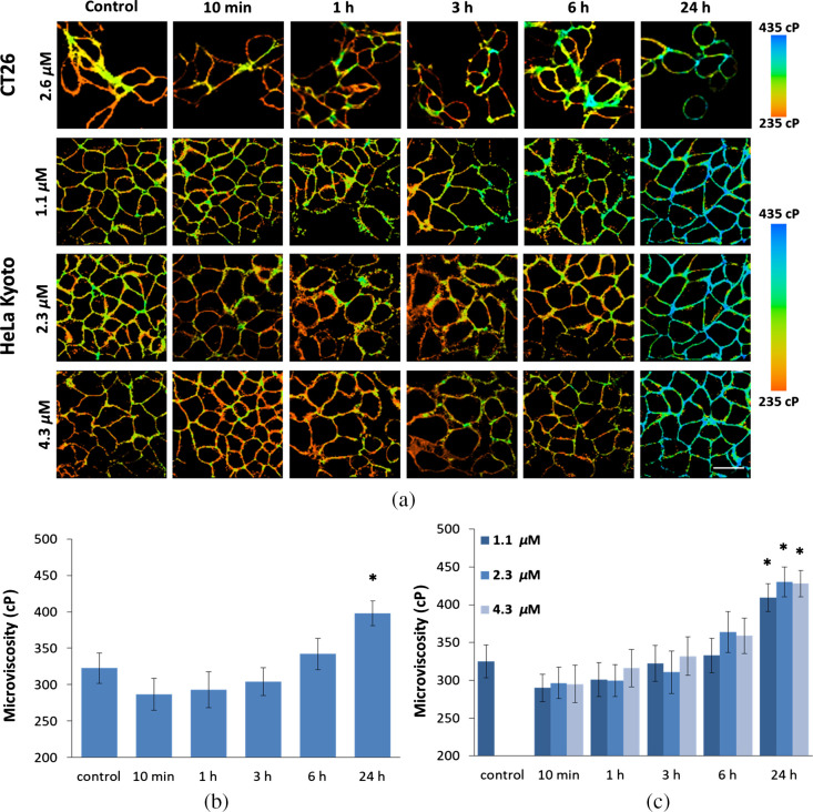
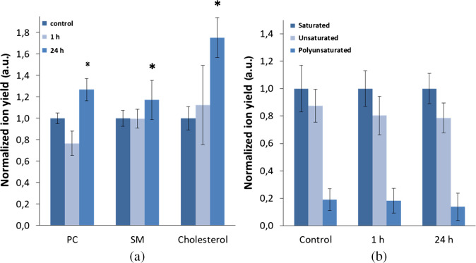
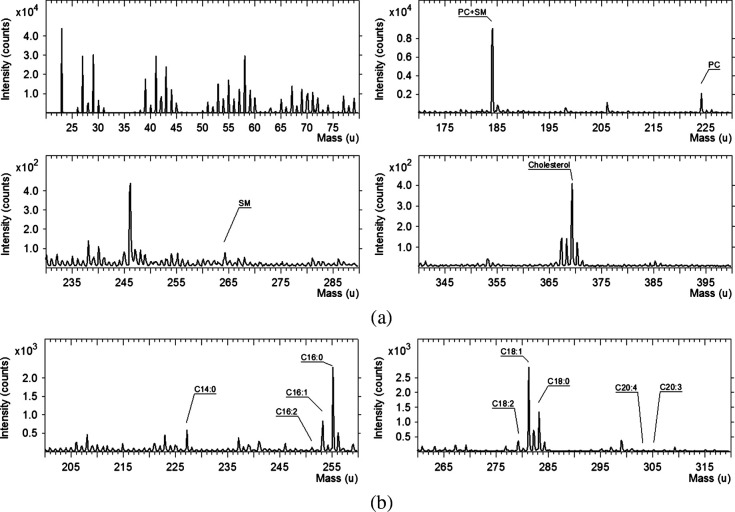
FLIM together with viscosity-sensitive boron dipyrromethene-based fluorescent molecular rotor was used to map the fluidity of the cell's membrane. Chemical analysis of membrane lipid composition was performed with time-of-flight secondary ion mass spectrometry (ToF-SIMS).
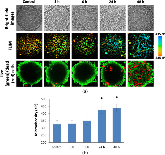
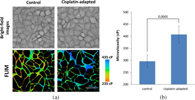
We detected a significant steady increase in membrane viscosity in viable cancer cells, both in cell monolayers and tumor spheroids, upon prolonged treatment with cisplatin, as well as in cisplatin-adapted cell line. ToF-SIMS revealed correlative changes in lipid profile of cisplatin-treated cells.
These results suggest an involvement of membrane viscosity in the cell adaptation to the drug and in the acquisition of drug resistance.
尽管细胞膜在调节药物活性方面很重要,但药物治疗对其物理性质的影响仍知之甚少。荧光寿命成像显微镜(FLIM)与特定粘度敏感荧光分子转子的结合,允许以高时空分辨率定量测量细胞膜的粘度,甚至可以测量到单个细胞细胞器。
我们的工作旨在使用 FLIM 分析顺铂化疗过程中活癌细胞的质膜微粘度,并将观察到的变化与脂质组成和细胞对治疗的反应相关联。
FLIM 与粘度敏感的硼二吡咯甲川基荧光分子转子一起用于绘制细胞膜流动性图。采用飞行时间二次离子质谱(ToF-SIMS)对膜脂质组成进行化学分析。
我们在顺铂处理的活癌细胞(包括细胞单层和肿瘤球体)中检测到膜粘度的持续显著增加,并且在顺铂适应细胞系中也观察到了这种增加。ToF-SIMS 揭示了顺铂处理细胞的脂质谱的相关变化。
这些结果表明,膜粘度参与了细胞对药物的适应和获得耐药性的过程。