Faculty of Biological and Environmental Sciences, Molecular and Integrative Biosciences, University of Helsinki, Helsinki, Finland.
Neuroscience Center (HiLIFE), University of Helsinki, Helsinki, Finland.
Epilepsia. 2021 Apr;62(4):908-919. doi: 10.1111/epi.16790. Epub 2020 Dec 18.
Birth asphyxia (BA) is often associated with seizures that may exacerbate the ensuing hypoxic-ischemic encephalopathy. In rodent models of BA, exposure to hypoxia is used to evoke seizures, that commence already during the insult. This is in stark contrast to clinical BA, in which seizures are typically seen upon recovery. Here, we introduce a term-equivalent rat model of BA, in which seizures are triggered after exposure to asphyxia.
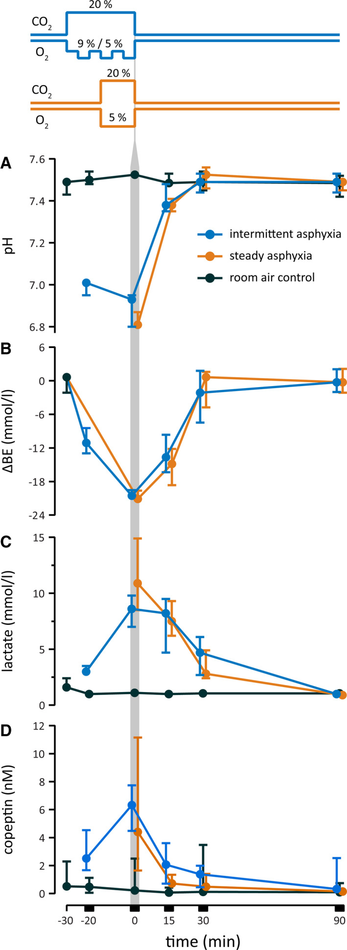
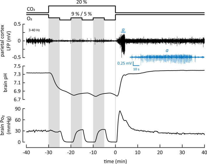
Postnatal day 11-12 male rat pups were exposed to steady asphyxia (15 min; air containing 5% O + 20% CO ) or to intermittent asphyxia (30 min; three 5 + 5-min cycles of 9% and 5% O at 20% CO ). Cortical activity and electrographic seizures were recorded in freely behaving animals. Simultaneous electrode measurements of intracortical pH, Po , and local field potentials (LFPs) were made under urethane anesthesia.
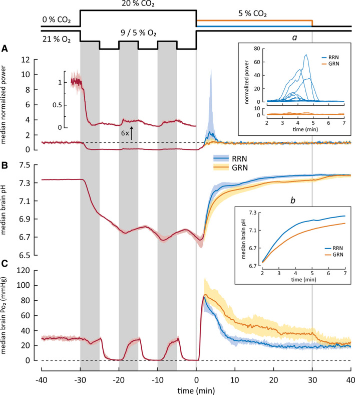
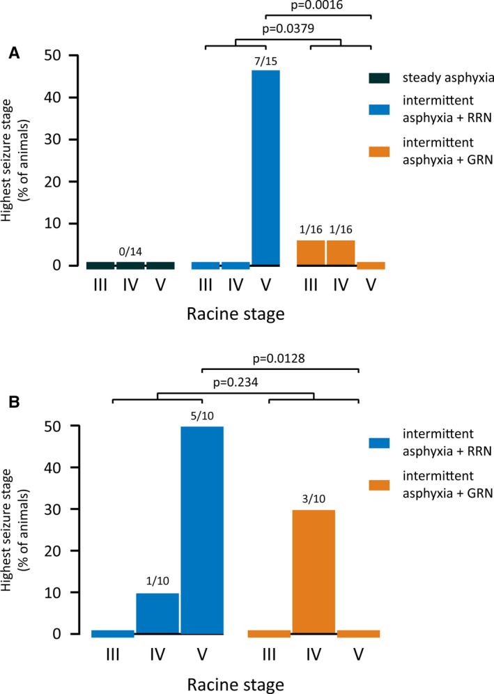
Both protocols decreased blood pH to <7.0 and brain pH from 7.3 to 6.7 and led to a fall in base excess by 20 mmol·L . Electrographic seizures with convulsions spanning the entire Racine scale were triggered after intermittent but not steady asphyxia. In the presence of 20% CO , brain Po was only transiently affected by 9% ambient O but fell below detection level during the steps to 5% O , and LFP activity was nearly abolished. Post-asphyxia seizures were strongly suppressed when brain pH recovery was slowed down by 5% CO .
The rate of brain pH recovery has a strong influence on post-asphyxia seizure propensity. The recurring hypoxic episodes during intermittent asphyxia promote neuronal excitability, which leads to seizures only after the suppressing effect of the hypercapnic acidosis is relieved. The present rodent model of BA is to our best knowledge the first one in which, consistent with clinical BA, behavioral and electrographic seizures are triggered after and not during the BA-mimicking insult.
出生窒息(BA)常伴有癫痫发作,可能会加重随后的缺氧缺血性脑病。在 BA 的啮齿动物模型中,使用缺氧来诱发癫痫发作,这些发作在损伤过程中已经开始。这与临床 BA 形成鲜明对比,在临床 BA 中,癫痫发作通常在恢复后出现。在这里,我们引入了一种与 BA 术语相当的大鼠模型,其中癫痫发作是在窒息后触发的。
在 11-12 日龄雄性大鼠幼崽出生后,将其暴露于持续窒息(15 分钟;含 5%O 的空气+20%CO )或间歇性窒息(30 分钟;三个 9%和 5%O 的 5+5 分钟循环,在 20%CO 下)。在自由活动的动物中记录皮质活动和脑电图癫痫发作。在 1% 戊巴比妥钠麻醉下同时进行皮质内 pH、Po 和局部场电位(LFP)的电极测量。
两种方案均使血 pH 值降至<7.0,脑 pH 值从 7.3 降至 6.7,并使基础不足降低 20mmol·L 。间歇性而非持续性窒息后会引发具有全身抽搐的电癫痫发作。在 20%CO 存在的情况下,脑 Po 仅被 9%环境 O 短暂影响,但在降至 5%O 的过程中降至检测水平以下,而 LFP 活动几乎被消除。当脑 pH 值恢复因 5%CO 而减慢时,窒息后癫痫发作受到强烈抑制。
脑 pH 值恢复的速度对窒息后癫痫发作的倾向有很大影响。间歇性窒息过程中反复出现的缺氧事件会促进神经元兴奋性,只有在高碳酸酸中毒的抑制作用解除后才会引发癫痫发作。据我们所知,本 BA 啮齿动物模型是第一个与临床 BA 一致的模型,即在模仿 BA 的损伤后而不是在损伤过程中引发行为和脑电图癫痫发作。