• 文献检索
  • 文档翻译
  • 深度研究
  • 学术资讯
  • Suppr Zotero 插件Zotero 插件
  • 邀请有礼
  • 套餐&价格
  • 历史记录
应用&插件
Suppr Zotero 插件Zotero 插件浏览器插件Mac 客户端Windows 客户端微信小程序
定价
高级版会员购买积分包购买API积分包
服务
文献检索文档翻译深度研究API 文档MCP 服务
关于我们
关于 Suppr公司介绍联系我们用户协议隐私条款
关注我们

Suppr 超能文献

核心技术专利:CN118964589B侵权必究
粤ICP备2023148730 号-1Suppr @ 2026

文献检索

告别复杂PubMed语法,用中文像聊天一样搜索,搜遍4000万医学文献。AI智能推荐,让科研检索更轻松。

立即免费搜索

文件翻译

保留排版,准确专业,支持PDF/Word/PPT等文件格式,支持 12+语言互译。

免费翻译文档

深度研究

AI帮你快速写综述,25分钟生成高质量综述,智能提取关键信息,辅助科研写作。

立即免费体验

非人灵长类动物严重缺氧缺血性脑病模型相关的局灶性脑损伤

Focal Brain Injury Associated with a Model of Severe Hypoxic-Ischemic Encephalopathy in Nonhuman Primates.

作者信息

McAdams Ryan M, McPherson Ronald J, Kapur Raj P, Juul Sandra E

机构信息

Division of Neonatology, Department of Pediatrics, University of Washington, Seattle, WA, USA.

出版信息

Dev Neurosci. 2017;39(1-4):107-123. doi: 10.1159/000456658. Epub 2017 Mar 25.

DOI:10.1159/000456658
PMID:28343228
原文链接:https://pmc.ncbi.nlm.nih.gov/articles/PMC5519439/
Abstract

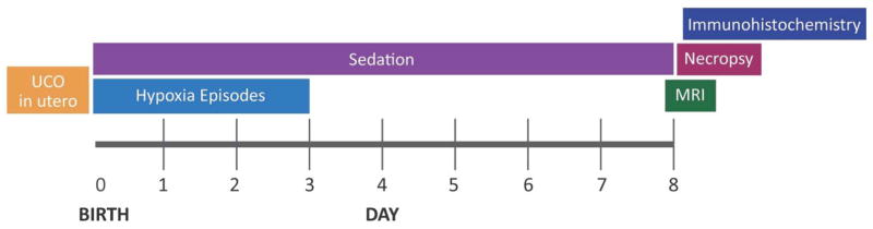
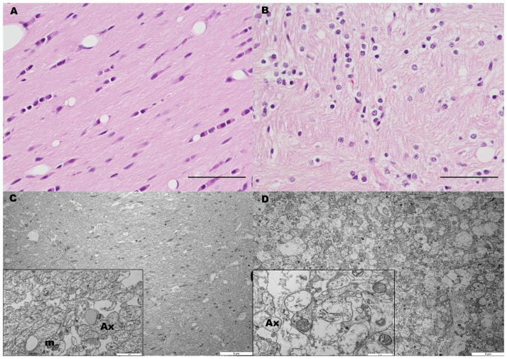
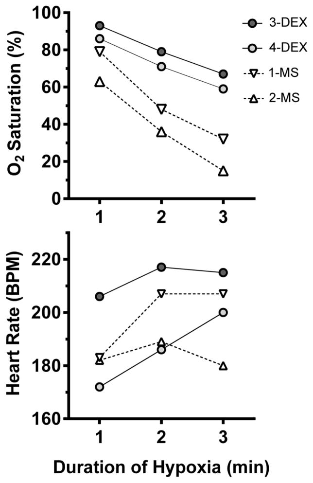
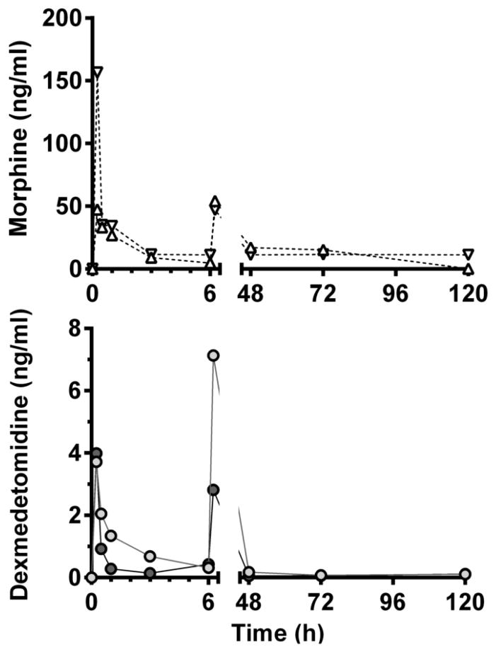
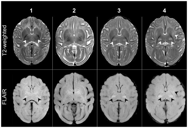
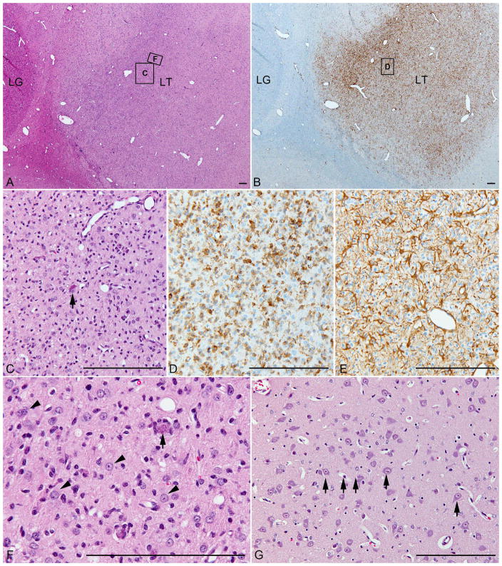
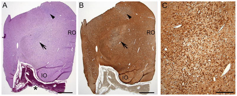
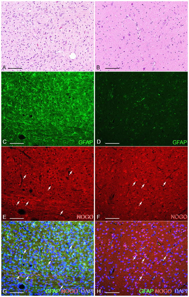
Worldwide, hypoxic-ischemic encephalopathy (HIE) is a major cause of neonatal mortality and morbidity. To better understand the mechanisms contributing to brain injury and improve outcomes in neonates with HIE, better preclinical animal models that mimic the clinical situation following birth asphyxia in term newborns are needed. In an effort to achieve this goal, we modified our nonhuman primate model of HIE induced by in utero umbilical cord occlusion (UCO) to include postnatal hypoxic episodes, in order to simulate apneic events in human neonates with HIE. We describe a cohort of 4 near-term fetal Macaca nemestrina that underwent 18 min of in utero UCO, followed by cesarean section delivery, resuscitation, and subsequent postnatal mechanical ventilation, with exposure to intermittent daily hypoxia (3 min, 8% O2 3-8 times daily for 3 days). After delivery, all animals demonstrated severe metabolic acidosis (pH 7 ± 0.12; mean ± SD) and low APGAR scores (<5 at 10 min of age). Three of 4 animals had both electrographic and clinical seizures. Serial blood samples were collected and plasma metabolites were determined by 2-dimensional gas chromatography coupled with time-of-flight mass spectrometry (GC × GC-TOFMS). The 4 UCO animals and a single nonasphyxiated animal (delivered by cesarean section but without exposure to UCO or prolonged sedation) underwent brain magnetic resonance imaging (MRI) on day 8 of life. Thalamic injury was present on MRI in 3 UCO animals, but not in the control animal. Following necropsy on day 8, brain histopathology revealed neuronal injury/loss and gliosis in portions of the ventrolateral thalamus in all 4 UCO, with 2 animals also demonstrating putamen/globus pallidus involvement. In addition, all 4 UCO animals demonstrated brain stem gliosis, with neuronal loss present in the midbrain, pons, and lateral medulla in 3 of 4 animals. Transmission electron microscopy imaging of the brain tissues was performed, which demonstrated ultrastructural white matter abnormalities, characterized by perinuclear vacuolation and axonal dilation, in 3 of 4 animals. Immunolabeling of Nogo-A, a negative regulator of neuronal growth, was not increased in the injured brains compared to 2 control animals. Using GC × GC-TOFMS, we identified metabolites previously recognized as potential biomarkers of perinatal asphyxia. The basal ganglia-thalamus-brain stem injury produced by UCO is consistent with the deep nuclear/brainstem injury pattern seen in human neonates after severe, abrupt hypoxic-ischemic insults. The UCO model permits timely detection of biomarkers associated with specific patterns of neonatal brain injury, and it may ultimately be useful for validating therapeutic strategies to treat neonatal HIE.

摘要

在全球范围内,缺氧缺血性脑病(HIE)是新生儿死亡和发病的主要原因。为了更好地理解导致脑损伤的机制并改善HIE新生儿的预后,需要更好的临床前动物模型来模拟足月儿出生窒息后的临床情况。为了实现这一目标,我们对子宫内脐带闭塞(UCO)诱导的HIE非人灵长类动物模型进行了改良,使其包括出生后的缺氧发作,以模拟患有HIE的人类新生儿的呼吸暂停事件。我们描述了一组4只近足月胎儿猕猴,它们经历了18分钟的子宫内UCO,随后进行剖宫产分娩、复苏,以及随后的出生后机械通气,并暴露于间歇性每日缺氧(3分钟,8%氧气,每天3 - 8次,持续3天)。分娩后,所有动物均表现出严重代谢性酸中毒(pH 7 ± 0.12;平均值±标准差)和低阿氏评分(10分钟龄时<5分)。4只动物中有3只出现脑电图和临床癫痫发作。采集系列血样,并通过二维气相色谱与飞行时间质谱联用(GC×GC - TOFMS)测定血浆代谢物。4只UCO动物和1只未窒息动物(通过剖宫产分娩,但未暴露于UCO或长时间镇静)在出生后第8天进行了脑磁共振成像(MRI)。3只UCO动物的MRI显示丘脑损伤,而对照动物未出现。在出生后第8天尸检后,脑组织病理学显示所有4只UCO动物的腹外侧丘脑部分存在神经元损伤/丢失和胶质细胞增生,2只动物还表现出壳核/苍白球受累。此外,所有4只UCO动物均表现出脑干胶质细胞增生,4只动物中有3只在中脑、脑桥和延髓外侧出现神经元丢失。对脑组织进行了透射电子显微镜成像,4只动物中有3只显示超微结构白质异常,其特征为核周空泡化和轴突扩张。与2只对照动物相比,损伤脑内神经元生长的负调节因子Nogo - A的免疫标记未增加。使用GC×GC - TOFMS,我们鉴定出了先前被认为是围产期窒息潜在生物标志物的代谢物。UCO导致的基底神经节 - 丘脑 - 脑干损伤与人类新生儿在严重、突然缺氧缺血性损伤后所见的深部核团/脑干损伤模式一致。UCO模型能够及时检测与新生儿脑损伤特定模式相关的生物标志物,并且它最终可能有助于验证治疗新生儿HIE的治疗策略。

https://cdn.ncbi.nlm.nih.gov/pmc/blobs/9c95/5519439/3d013ea6c921/nihms851147f8.jpg
https://cdn.ncbi.nlm.nih.gov/pmc/blobs/9c95/5519439/d73b3683270f/nihms851147f1.jpg
https://cdn.ncbi.nlm.nih.gov/pmc/blobs/9c95/5519439/f0ce32173dca/nihms851147f2.jpg
https://cdn.ncbi.nlm.nih.gov/pmc/blobs/9c95/5519439/e5392a4fcf3d/nihms851147f3.jpg
https://cdn.ncbi.nlm.nih.gov/pmc/blobs/9c95/5519439/0fc1bd55024b/nihms851147f4.jpg
https://cdn.ncbi.nlm.nih.gov/pmc/blobs/9c95/5519439/9495303bbe64/nihms851147f5.jpg
https://cdn.ncbi.nlm.nih.gov/pmc/blobs/9c95/5519439/7f812ff95543/nihms851147f6.jpg
https://cdn.ncbi.nlm.nih.gov/pmc/blobs/9c95/5519439/7b7109324942/nihms851147f7.jpg
https://cdn.ncbi.nlm.nih.gov/pmc/blobs/9c95/5519439/3d013ea6c921/nihms851147f8.jpg

相似文献

1
Focal Brain Injury Associated with a Model of Severe Hypoxic-Ischemic Encephalopathy in Nonhuman Primates.非人灵长类动物严重缺氧缺血性脑病模型相关的局灶性脑损伤
Dev Neurosci. 2017;39(1-4):107-123. doi: 10.1159/000456658. Epub 2017 Mar 25.
2
Long-Term Neuropathological Changes Associated with Cerebral Palsy in a Nonhuman Primate Model of Hypoxic-Ischemic Encephalopathy.缺氧缺血性脑病非人灵长类动物模型中与脑瘫相关的长期神经病理学变化
Dev Neurosci. 2017;39(1-4):124-140. doi: 10.1159/000470903. Epub 2017 May 10.
3
Serial plasma metabolites following hypoxic-ischemic encephalopathy in a nonhuman primate model.非人类灵长类动物模型中缺氧缺血性脑病后的系列血浆代谢物
Dev Neurosci. 2015;37(2):161-71. doi: 10.1159/000370147. Epub 2015 Feb 27.
4
Concurrent erythropoietin and hypothermia treatment improve outcomes in a term nonhuman primate model of perinatal asphyxia.同时进行促红细胞生成素和低温治疗可改善围产期窒息的足月非人灵长类动物模型的预后。
Dev Neurosci. 2013;35(6):491-503. doi: 10.1159/000355460. Epub 2013 Nov 1.
5
Prenatal cord clamping in newborn Macaca nemestrina: a model of perinatal asphyxia.恒河猴新生儿产前脐带钳夹:围产期窒息模型
Dev Neurosci. 2007;29(4-5):311-20. doi: 10.1159/000105472.
6
Cytokine and chemokine responses to injury and treatment in a nonhuman primate model of hypoxic-ischemic encephalopathy treated with hypothermia and erythropoietin.细胞因子和趋化因子对接受低温和促红细胞生成素治疗的缺氧缺血性脑病非人灵长类动物模型的损伤和治疗的反应。
J Cereb Blood Flow Metab. 2021 Aug;41(8):2054-2066. doi: 10.1177/0271678X21991439. Epub 2021 Feb 7.
7
Development of epilepsy in newborns with moderate hypoxic-ischemic encephalopathy and neonatal seizures.中度缺氧缺血性脑病及新生儿惊厥的新生儿癫痫的发展
Brain Dev. 2009 Jan;31(1):64-8. doi: 10.1016/j.braindev.2008.04.001. Epub 2008 May 19.
8
Plasma vasopressin levels are closely associated with fetal hypotension and neuronal injury after hypoxia-ischemia in near-term fetal sheep.血浆血管加压素水平与近足月胎儿缺氧缺血后低血压和神经元损伤密切相关。
Pediatr Res. 2020 Dec;88(6):857-864. doi: 10.1038/s41390-020-0845-2. Epub 2020 Mar 17.
9
Diffusion Tensor Imaging Detects Occult Cerebellar Injury in Severe Neonatal Hypoxic-Ischemic Encephalopathy.弥散张量成像检测重度新生儿缺氧缺血性脑病中的隐匿性小脑损伤。
Dev Neurosci. 2017;39(1-4):207-214. doi: 10.1159/000454856. Epub 2017 Jan 18.
10
Endogenous hypothermic response to hypoxia reduces brain injury: Implications for modeling hypoxic-ischemic encephalopathy and therapeutic hypothermia in neonatal mice.内源性低温反应对缺氧的减少脑损伤:对新生儿缺氧缺血性脑病模型和治疗性低温的影响。
Exp Neurol. 2016 Sep;283(Pt A):264-75. doi: 10.1016/j.expneurol.2016.06.024. Epub 2016 Jun 25.

引用本文的文献

1
Research progress of neonatal hypoxic-ischemic encephalopathy in nonhuman primate models.非人灵长类动物模型中新生儿缺氧缺血性脑病的研究进展
Ibrain. 2023 Mar 26;9(2):183-194. doi: 10.1002/ibra.12097. eCollection 2023 Summer.
2
Regulation of glutamate transport and neuroinflammation in a term newborn rat model of hypoxic-ischaemic brain injury.足月新生大鼠缺氧缺血性脑损伤模型中谷氨酸转运与神经炎症的调节
Brain Neurosci Adv. 2022 May 20;6:23982128221097568. doi: 10.1177/23982128221097568. eCollection 2022 Jan-Dec.
3
Defining Longer-Term Outcomes in an Ovine Model of Moderate Perinatal Hypoxia-Ischemia.

本文引用的文献

1
Diffusion Tensor Imaging Abnormalities in the Cerebral White Matter Correlate with Sex-Dependent Neurobehavioral Deficits in Adult Mice with Neonatal Ischemia.脑白质扩散张量成像异常与新生儿缺血成年小鼠的性别依赖性神经行为缺陷相关。
Dev Neurosci. 2016;38(2):83-95. doi: 10.1159/000442943. Epub 2016 Mar 16.
2
The Effect of Perinatal Hypoxic/Ischemic Injury on Tyrosine Hydroxylase Expression in the Locus Coeruleus of the Human Neonate.围产期缺氧/缺血性损伤对人类新生儿蓝斑中酪氨酸羟化酶表达的影响。
Dev Neurosci. 2016;38(1):41-53. doi: 10.1159/000439270. Epub 2015 Dec 9.
3
Effects of Neonatal Hypoxic-Ischemic Injury and Hypothermic Neuroprotection on Neural Progenitor Cells in the Mouse Hippocampus.
在绵羊围产期中度缺氧缺血模型中定义长期结局。
Dev Neurosci. 2022;44(4-5):277-294. doi: 10.1159/000525150. Epub 2022 May 19.
4
Pathophysiology of Perinatal Asphyxia in Humans and Animal Models.人类和动物模型中围产期窒息的病理生理学
Biomedicines. 2022 Feb 1;10(2):347. doi: 10.3390/biomedicines10020347.
5
Neuroimaging in the term newborn with neonatal encephalopathy.足月新生儿脑病的神经影像学。
Semin Fetal Neonatal Med. 2021 Oct;26(5):101304. doi: 10.1016/j.siny.2021.101304. Epub 2021 Oct 29.
6
A Metabolomic Approach in Search of Neurobiomarkers of Perinatal Asphyxia: A Review of the Current Literature.一种寻找围产期窒息神经生物标志物的代谢组学方法:当前文献综述
Front Pediatr. 2021 Jun 25;9:674585. doi: 10.3389/fped.2021.674585. eCollection 2021.
7
Integrating neuroimaging biomarkers into the multicentre, high-dose erythropoietin for asphyxia and encephalopathy (HEAL) trial: rationale, protocol and harmonisation.将神经影像学生物标志物纳入多中心高剂量促红细胞生成素治疗窒息和脑病(HEAL)试验:原理、方案与协调
BMJ Open. 2021 Apr 22;11(4):e043852. doi: 10.1136/bmjopen-2020-043852.
8
Metabolomic analysis and mass spectrometry imaging after neonatal stroke and cell therapies in mouse brains.新生儿脑卒中和细胞治疗后的代谢组学分析和质谱成像。
Sci Rep. 2020 Dec 14;10(1):21881. doi: 10.1038/s41598-020-78930-x.
9
Exploring Perinatal Asphyxia by Metabolomics.通过代谢组学探索围产期窒息
Metabolites. 2020 Apr 4;10(4):141. doi: 10.3390/metabo10040141.
10
Electroencephalogram studies of hypoxic ischemia in fetal and neonatal animal models.胎儿和新生儿动物模型中缺氧缺血的脑电图研究。
Neural Regen Res. 2020 May;15(5):828-837. doi: 10.4103/1673-5374.268892.
新生儿缺氧缺血性损伤及低温神经保护对小鼠海马神经祖细胞的影响
Dev Neurosci. 2015;37(4-5):428-39. doi: 10.1159/000430862. Epub 2015 Jun 13.
4
Dexmedetomidine reduces cranial temperature in hypothermic neonatal rats.右美托咪定可降低低温新生大鼠的颅温。
Pediatr Res. 2015 Jun;77(6):772-8. doi: 10.1038/pr.2015.45. Epub 2015 Mar 9.
5
Executive summary: Neonatal encephalopathy and neurologic outcome, second edition. Report of the American College of Obstetricians and Gynecologists' Task Force on Neonatal Encephalopathy.执行摘要:新生儿脑病与神经学结局,第二版。美国妇产科医师学会新生儿脑病特别工作组报告。
Obstet Gynecol. 2014 Apr;123(4):896-901. doi: 10.1097/01.AOG.0000445580.65983.d2.
6
Nogo limits neural plasticity and recovery from injury.Nogo蛋白限制神经可塑性和损伤后的恢复。
Curr Opin Neurobiol. 2014 Aug;27:53-60. doi: 10.1016/j.conb.2014.02.011. Epub 2014 Mar 12.
7
Comparative primate neuroimaging: insights into human brain evolution.比较灵长类神经影像学:对人类大脑进化的洞察。
Trends Cogn Sci. 2014 Jan;18(1):46-55. doi: 10.1016/j.tics.2013.09.013.
8
Concurrent erythropoietin and hypothermia treatment improve outcomes in a term nonhuman primate model of perinatal asphyxia.同时进行促红细胞生成素和低温治疗可改善围产期窒息的足月非人灵长类动物模型的预后。
Dev Neurosci. 2013;35(6):491-503. doi: 10.1159/000355460. Epub 2013 Nov 1.
9
Four decades of leading-edge research in the reproductive and developmental sciences: the Infant Primate Research Laboratory at the University of Washington National Primate Research Center.四十年在生殖和发育科学方面的前沿研究:华盛顿大学国家灵长类动物研究中心的婴儿灵长类动物研究实验室。
Am J Primatol. 2013 Nov;75(11):1063-83. doi: 10.1002/ajp.22175. Epub 2013 Jul 19.
10
Nogo-A represses anatomical and synaptic plasticity in the central nervous system.Nogo-A 抑制中枢神经系统的解剖和突触可塑性。
Physiology (Bethesda). 2013 May;28(3):151-63. doi: 10.1152/physiol.00052.2012.