Department of Biomedical Engineering, Faculty of Environment and Life, Beijing University of Technology, Beijing, China.
Julius Wolff Institute and BIH Center for Regenerative Therapies, Berlin Institute of Health and Charité-Universitätsmedizin Berlin, Berlin, Germany.
Sci Rep. 2020 Dec 18;10(1):22299. doi: 10.1038/s41598-020-79098-0.
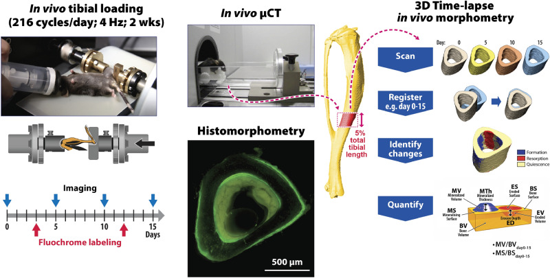
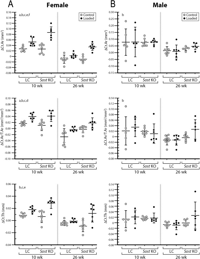
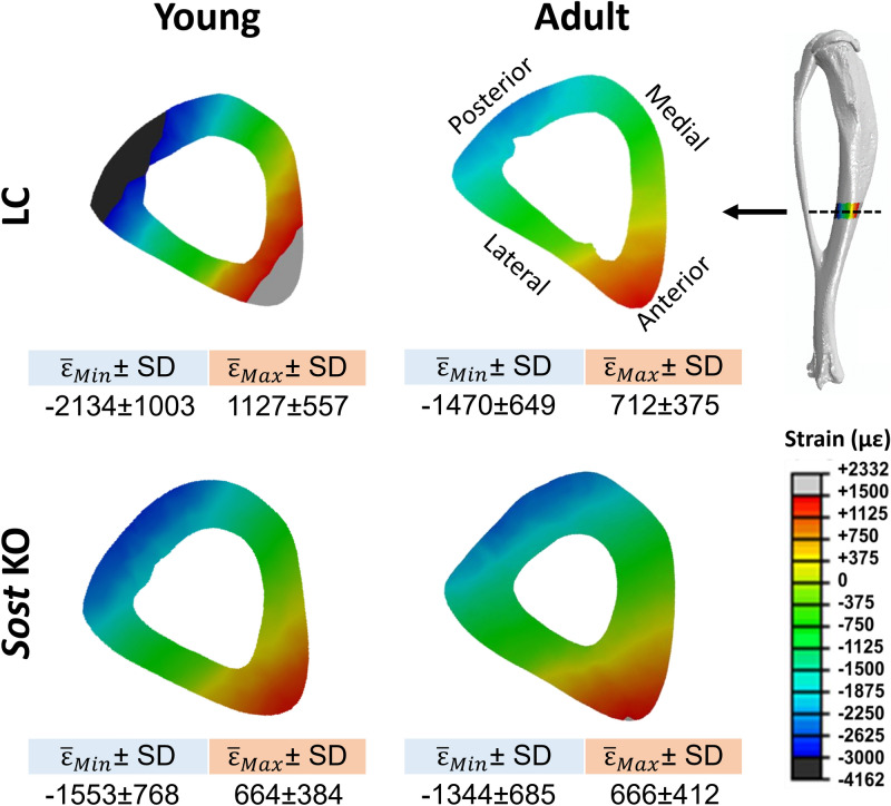
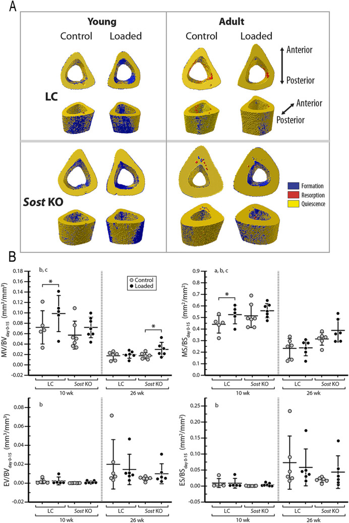
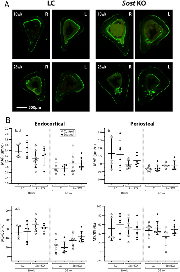
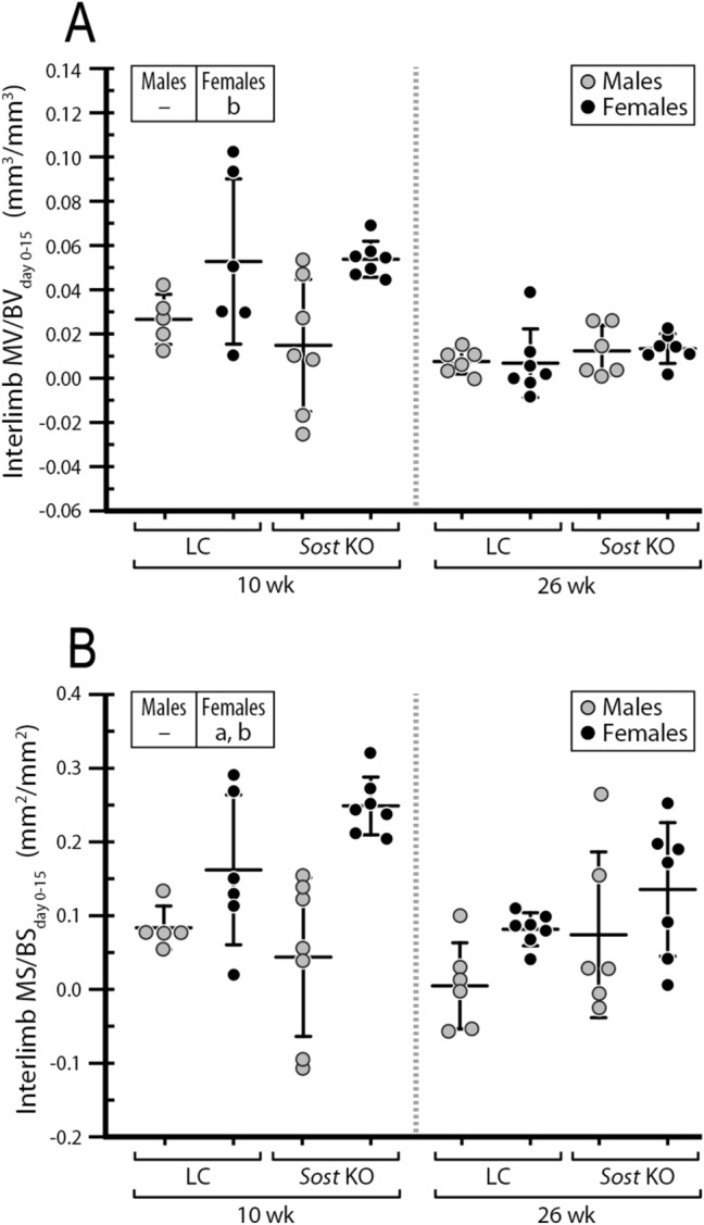
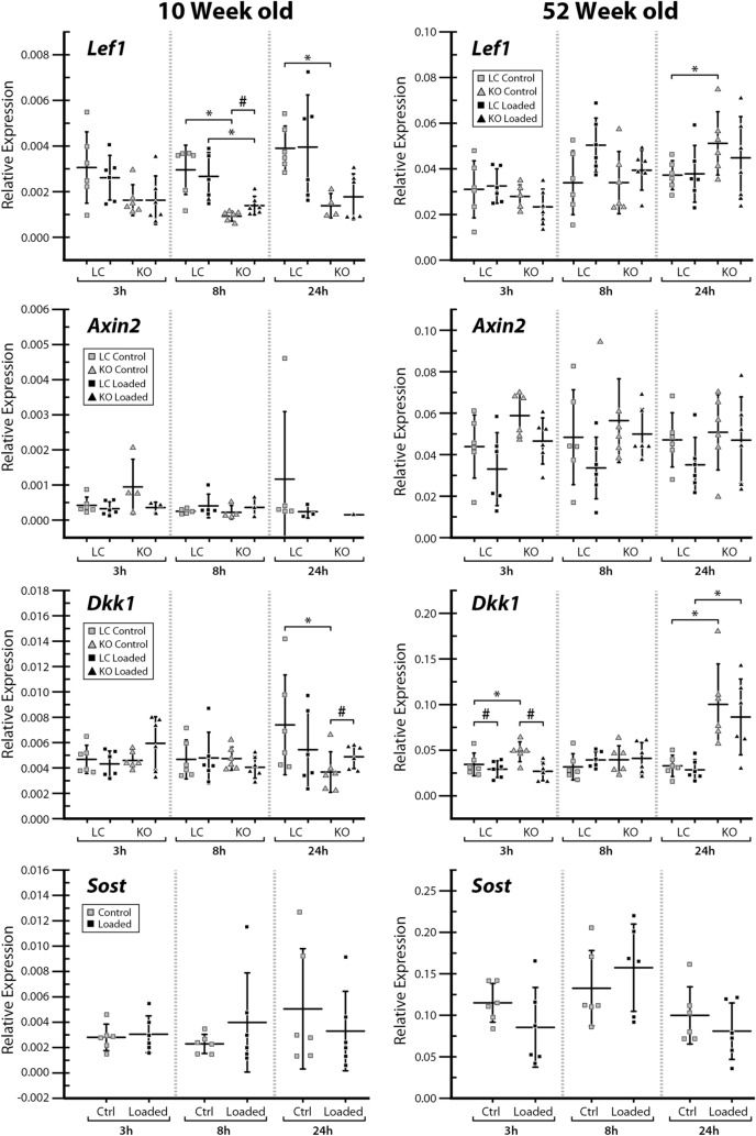
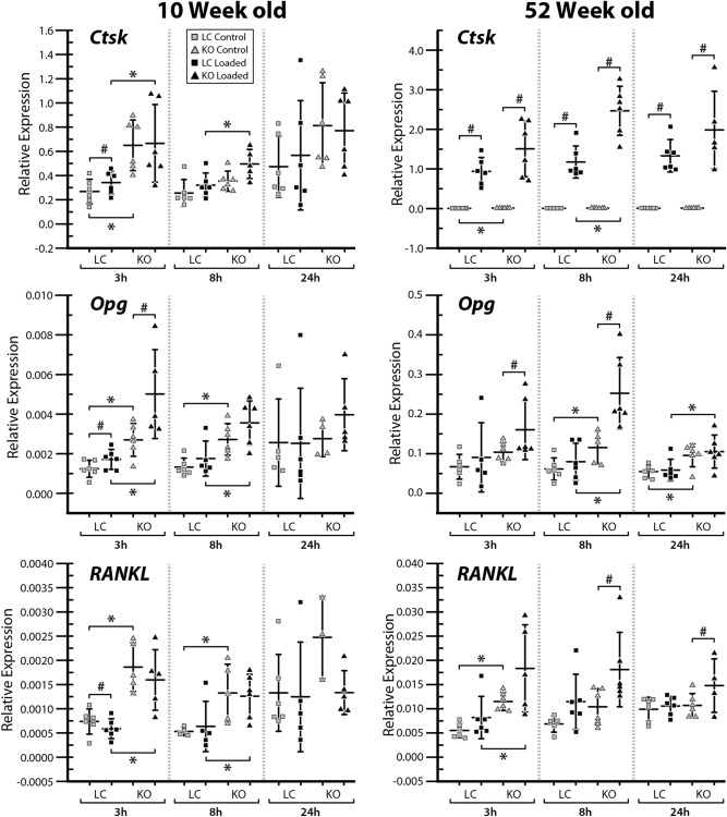
Loss-of-function mutations in the Sost gene lead to high bone mass phenotypes. Pharmacological inhibition of Sost/sclerostin provides a new drug strategy for treating osteoporosis. Questions remain as to how physical activity may affect bone mass under sclerostin inhibition and if that effect differs between males and females. We previously observed in female Sost knockout (KO) mice an enhanced cortical bone formation response to a moderate level of applied loading (900 με at the tibial midshaft). The purpose of the present study was to examine cortical bone adaptation to the same strain level applied to male Sost KO mice. Strain-matched in vivo compressive loading was applied to the tibiae of 10-, 26- and 52-week-old male Sost KO and littermate control (LC) mice. The effect of tibial loading on bone (re)modeling was measured by microCT, 3D time-lapse in vivo morphometry, 2D histomorphometry and gene expression analyses. As expected, Sost deficiency led to high cortical bone mass in 10- and 26-week-old male mice as a result of increased bone formation. However, the enhanced bone formation associated with Sost deficiency did not appear to diminish with skeletal maturation. An increase in bone resorption was observed with skeletal maturation in male LC and Sost KO mice. Two weeks of in vivo loading (900 με at the tibial midshaft) induced only a mild anabolic response in 10- and 26-week-old male mice, independent of Sost deficiency. A decrease in the Wnt inhibitor Dkk1 expression was observed 3 h after loading in 52-week-old Sost KO and LC mice, and an increase in Lef1 expression was observed 8 h after loading in 10-week-old Sost KO mice. The current results suggest that long-term inhibition of sclerostin in male mice does not influence the adaptive response of cortical bone to moderate levels of loading. In contrast with our previous strain-matched study in females showing enhanced bone responses with Sost ablation, these results in males indicate that the influence of Sost deficiency on the cortical bone formation response to a moderate level of loading differs between males and females. Clinical studies examining antibodies to inhibit sclerostin may need to consider that the efficacy of additional physical activity regimens may be sex dependent.
Sost 基因的功能丧失突变导致高骨量表型。Sost/sclerostin 的药理学抑制为治疗骨质疏松症提供了一种新的药物策略。目前仍存在疑问的是,在抑制 Sost 后,体力活动如何影响骨量,以及这种影响在男性和女性之间是否存在差异。我们之前在雌性 Sost 敲除 (KO) 小鼠中观察到,在胫骨中段施加适度水平的应用负载(900 με)时,皮质骨形成反应增强。本研究的目的是检查相同应变水平施加于雄性 Sost KO 小鼠时皮质骨的适应性。对 10、26 和 52 周龄雄性 Sost KO 和同窝对照 (LC) 小鼠的胫骨进行应变匹配的体内压缩加载。通过 microCT、3D 时间延迟活体形态计量学、2D 组织形态计量学和基因表达分析来测量胫骨加载对骨(再)建模的影响。正如预期的那样,由于骨形成增加,Sost 缺乏导致 10 至 26 周龄雄性小鼠的皮质骨量增加。然而,与骨骼成熟相关的 Sost 缺乏引起的骨形成增加似乎并没有随着骨骼成熟而减少。在雄性 LC 和 Sost KO 小鼠中,随着骨骼成熟,观察到骨吸收增加。在 10 至 26 周龄雄性小鼠中,2 周的体内加载(胫骨中段 900 με)独立于 Sost 缺乏,仅引起轻度的合成代谢反应。在 52 周龄 Sost KO 和 LC 小鼠中,加载后 3 小时观察到 Wnt 抑制剂 Dkk1 表达下降,在 10 周龄 Sost KO 小鼠中,加载后 8 小时观察到 Lef1 表达增加。目前的结果表明,长期抑制雄性小鼠的 Sostin 并不影响皮质骨对适度负荷的适应性反应。与我们之前在雌性中的研究结果不同,该研究表明,Sost 缺失对适度负荷下皮质骨形成反应的影响在男性和女性之间存在差异。检查抑制 Sost 抗体的临床研究可能需要考虑,额外的体育活动方案的疗效可能取决于性别。