• 文献检索
  • 文档翻译
  • 深度研究
  • 学术资讯
  • Suppr Zotero 插件Zotero 插件
  • 邀请有礼
  • 套餐&价格
  • 历史记录
应用&插件
Suppr Zotero 插件Zotero 插件浏览器插件Mac 客户端Windows 客户端微信小程序
定价
高级版会员购买积分包购买API积分包
服务
文献检索文档翻译深度研究API 文档MCP 服务
关于我们
关于 Suppr公司介绍联系我们用户协议隐私条款
关注我们

Suppr 超能文献

核心技术专利:CN118964589B侵权必究
粤ICP备2023148730 号-1Suppr @ 2026

文献检索

告别复杂PubMed语法,用中文像聊天一样搜索,搜遍4000万医学文献。AI智能推荐,让科研检索更轻松。

立即免费搜索

文件翻译

保留排版,准确专业,支持PDF/Word/PPT等文件格式,支持 12+语言互译。

免费翻译文档

深度研究

AI帮你快速写综述,25分钟生成高质量综述,智能提取关键信息,辅助科研写作。

立即免费体验

眼轴长度对人眼视网膜神经节细胞层的影响。

The Influence of Axial Length Upon the Retinal Ganglion Cell Layer of the Human Eye.

机构信息

Department of Radiology, Scheie Eye Institute, Penn Presbyterian Medical Center, Philadelphia, PA, USA.

Department of Neurology, Scheie Eye Institute, Penn Presbyterian Medical Center, Philadelphia, PA, USA.

出版信息

Transl Vis Sci Technol. 2020 Dec 7;9(13):9. doi: 10.1167/tvst.9.13.9. eCollection 2020 Dec.

DOI:10.1167/tvst.9.13.9
PMID:33344053
原文链接:https://pmc.ncbi.nlm.nih.gov/articles/PMC7726585/
Abstract

PURPOSE

Variation in retinal thickness with eye size complicates efforts to estimate retinal ganglion cell number from optical coherence tomography (OCT) measures. We examined the relationship among axial length, the thickness and volume of the ganglion cell layer (GCL), and the size of the optic chiasm.

METHODS

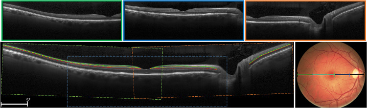
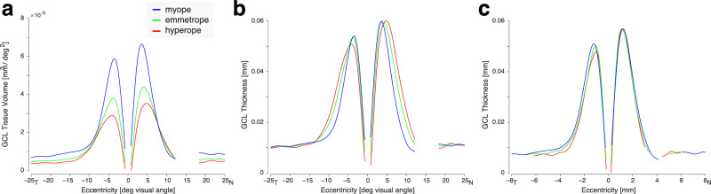
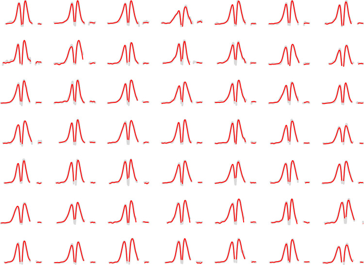
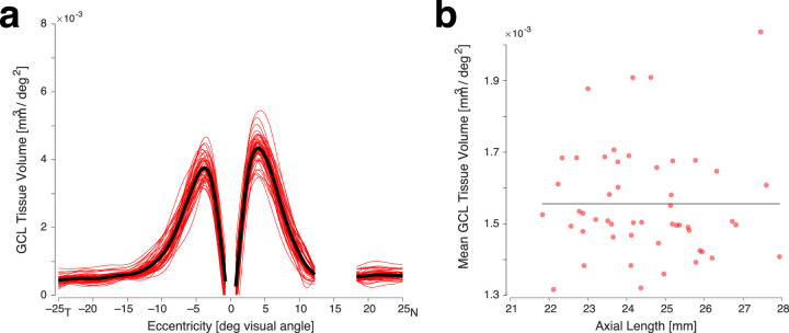
We used OCT to measure GCL thickness over 50 degrees of the horizontal meridian in 50 healthy participants with a wide range of axial lengths. Using a model eye informed by individual biometry, we converted GCL thickness to tissue volume per square degree. We also measured the volume of the optic chiasm for 40 participants using magnetic resonance imaging (MRI).

RESULTS

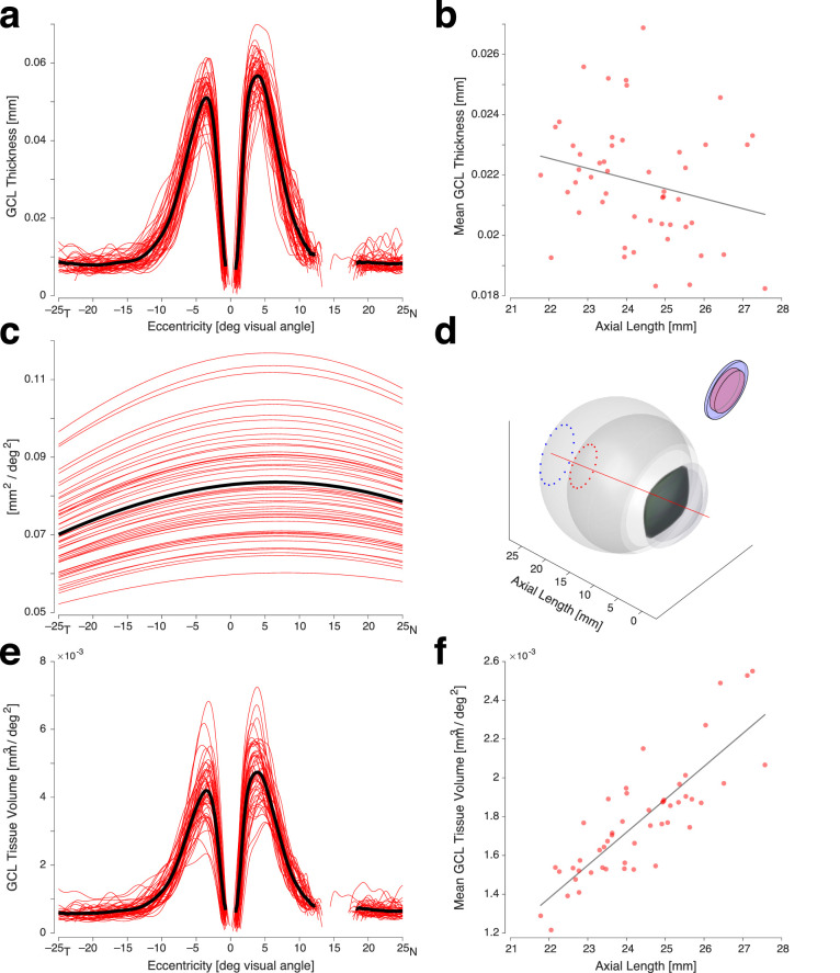
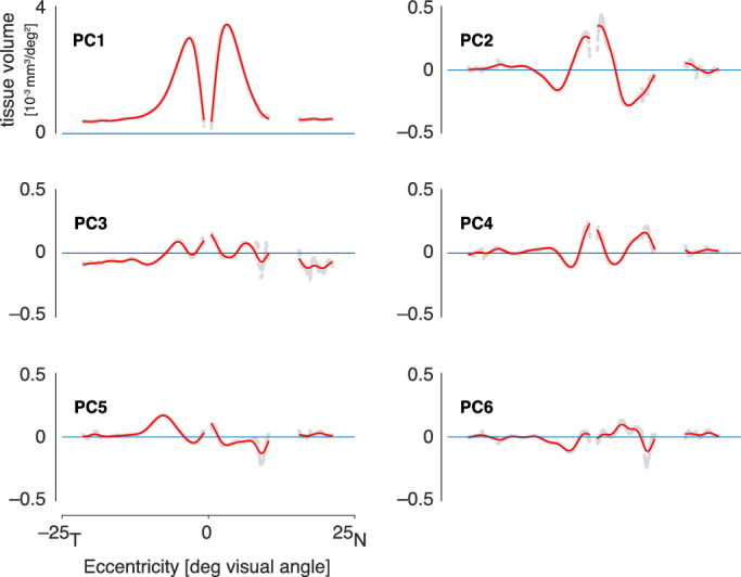
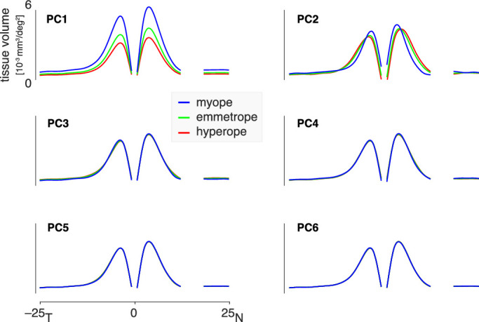
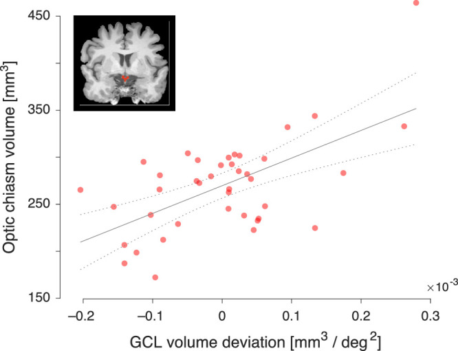
There is a positive relationship between GCL tissue volume and axial length. Given prior psychophysical results, we conclude that increased axial length is associated with increased retinal ganglion cell size, decreased cell packing, or both. We characterize how retinal ganglion cell tissue varies systematically in volume and spatial distribution as a function of axial length. This model allows us to remove the effect of axial length from individual difference measures of GCL volume. We find that variation in this adjusted GCL volume correlates well with the size of the optic chiasm.

CONCLUSIONS

Our results provide the volume of ganglion cell tissue in the retina, adjusted for the presumed effects of axial length upon ganglion cell size and/or packing. The resulting volume measure accounts for individual differences in the size of the optic chiasm, supporting its use to characterize the post-retinal visual pathway.

TRANSLATIONAL RELEVANCE

Variations in ametropia can confound clinical measures of retinal features. We present a framework within which the thickness and volume of retinal structures can be measured and corrected for the effects of axial length.

摘要

目的

眼球尺寸的变化会使从光学相干断层扫描(OCT)测量结果估计视网膜神经节细胞数量的工作变得复杂。我们研究了眼轴长度、神经节细胞层(GCL)的厚度和体积以及视交叉大小之间的关系。

方法

我们使用 OCT 在 50 名具有广泛眼轴长度的健康参与者的水平子午线的 50 度范围内测量 GCL 厚度。使用个体生物测量学提供的模型眼,我们将 GCL 厚度转换为每平方度的组织体积。我们还使用磁共振成像(MRI)测量了 40 名参与者的视交叉体积。

结果

GCL 组织体积与眼轴长度呈正相关。鉴于先前的心理物理学结果,我们得出结论,眼轴长度的增加与视网膜神经节细胞大小的增加、细胞密度的降低或两者兼有关。我们描述了视网膜神经节细胞组织如何作为眼轴长度的函数在体积和空间分布上系统地变化。该模型使我们能够从 GCL 体积的个体差异测量中去除眼轴长度的影响。我们发现,经调整的 GCL 体积的变化与视交叉的大小密切相关。

结论

我们的结果提供了视网膜神经节细胞组织的体积,该体积针对眼轴长度对神经节细胞大小和/或密度的假定影响进行了调整。所得的体积测量值考虑了视交叉大小的个体差异,支持其用于描述视网膜后视觉通路的特征。

翻译

本文描述了如何从视网膜神经节细胞层(GCL)的厚度和体积的 OCT 测量结果中去除眼轴长度的影响,并提供了一种方法来调整 GCL 体积,以考虑到视交叉大小的个体差异。该研究为研究和诊断与眼轴长度相关的眼部疾病提供了新的思路和方法。

https://cdn.ncbi.nlm.nih.gov/pmc/blobs/99c2/7726585/30e9df5d2943/tvst-9-13-9-f008.jpg
https://cdn.ncbi.nlm.nih.gov/pmc/blobs/99c2/7726585/bcd678384301/tvst-9-13-9-f001.jpg
https://cdn.ncbi.nlm.nih.gov/pmc/blobs/99c2/7726585/a5ff8f5e3f05/tvst-9-13-9-f002.jpg
https://cdn.ncbi.nlm.nih.gov/pmc/blobs/99c2/7726585/b391fb10989a/tvst-9-13-9-f003.jpg
https://cdn.ncbi.nlm.nih.gov/pmc/blobs/99c2/7726585/c342bb22de99/tvst-9-13-9-f004.jpg
https://cdn.ncbi.nlm.nih.gov/pmc/blobs/99c2/7726585/192cd870e819/tvst-9-13-9-f005.jpg
https://cdn.ncbi.nlm.nih.gov/pmc/blobs/99c2/7726585/8700bca7da4e/tvst-9-13-9-f006.jpg
https://cdn.ncbi.nlm.nih.gov/pmc/blobs/99c2/7726585/205f31ae07db/tvst-9-13-9-f007.jpg
https://cdn.ncbi.nlm.nih.gov/pmc/blobs/99c2/7726585/30e9df5d2943/tvst-9-13-9-f008.jpg

相似文献

1
The Influence of Axial Length Upon the Retinal Ganglion Cell Layer of the Human Eye.眼轴长度对人眼视网膜神经节细胞层的影响。
Transl Vis Sci Technol. 2020 Dec 7;9(13):9. doi: 10.1167/tvst.9.13.9. eCollection 2020 Dec.
2
Retinal ganglion cell layer thickness and volume measured by OCT changes with age, sex, and axial length in a healthy population.在健康人群中,OCT 测量的视网膜神经节细胞层厚度和体积随年龄、性别和眼轴长度而变化。
BMC Ophthalmol. 2022 Jun 24;22(1):278. doi: 10.1186/s12886-022-02488-7.
3
Thickness mapping of individual retinal layers and sectors by Spectralis SD-OCT in Autosomal Dominant Optic Atrophy.Spectralis SD-OCT 对常染色体显性视神经萎缩个体视网膜各层和各象限的厚度进行测绘。
Acta Ophthalmol. 2018 May;96(3):251-256. doi: 10.1111/aos.13588. Epub 2017 Nov 1.
4
Effects of axial length on retinal nerve fiber layer and macular ganglion cell-inner plexiform layer measured by spectral-domain OCT.轴向长度对通过频域光学相干断层扫描测量的视网膜神经纤维层和黄斑神经节细胞-内丛状层的影响。
Arq Bras Oftalmol. 2020 Aug;83(4):269-276. doi: 10.5935/0004-2749.20200039. Epub 2020 Jul 29.
5
[The correlation of ganglion cell layer thickness with visual field defect in non-functional pituitary adenoma with chiasm compression].[非功能性垂体腺瘤压迫视交叉时神经节细胞层厚度与视野缺损的相关性]
Zhonghua Yan Ke Za Zhi. 2019 Mar 11;55(3):186-194. doi: 10.3760/cma.j.issn.0412-4081.2019.03.007.
6
Effect of refractive errors/axial length on peripapillary retinal nerve fibre layer thickness (RNFL) measured by Topcon SD-OCT.屈光不正/眼轴长度对Topcon SD-OCT测量的视盘周围视网膜神经纤维层厚度(RNFL)的影响。
J Pak Med Assoc. 2018 Jul;68(7):1054-1059.
7
Determinants of normal retinal nerve fiber layer thickness measured by Stratus OCT.通过Stratus OCT测量的正常视网膜神经纤维层厚度的决定因素。
Ophthalmology. 2007 Jun;114(6):1046-52. doi: 10.1016/j.ophtha.2006.08.046. Epub 2007 Jan 8.
8
Regression Analysis of Optical Coherence Tomography Disc Variables for Glaucoma Diagnosis.用于青光眼诊断的光学相干断层扫描视盘变量的回归分析
J Glaucoma. 2016 Aug;25(8):634-42. doi: 10.1097/IJG.0000000000000378.
9
Determinants of perimacular inner retinal layer thickness in normal eyes measured by Fourier-domain optical coherence tomography.频域光相干断层扫描测量正常眼周边黄斑内层视网膜厚度的影响因素。
Invest Ophthalmol Vis Sci. 2011 May 18;52(6):3413-8. doi: 10.1167/iovs.10-6648.
10
Retinal nerve fibre and ganglion cell inner plexiform layer analysis by optical coherence tomography in asymptomatic empty sella patients.无症状空泡蝶鞍患者的光学相干断层扫描分析视网膜神经纤维和节细胞内丛状层。
Int J Neurosci. 2020 Jan;130(1):45-51. doi: 10.1080/00207454.2019.1660328. Epub 2019 Sep 13.

引用本文的文献

1
Sustained Experimental Myopia Exacerbates the Effect of Eye Growth on Retinal Ganglion Cell Density and Function.持续性实验性近视会加剧眼球生长对视网膜神经节细胞密度和功能的影响。
Int J Mol Sci. 2025 Mar 20;26(6):2824. doi: 10.3390/ijms26062824.
2
Anatomical Features can Affect OCT Measures Used for Clinical Decisions and Clinical Trial Endpoints.解剖学特征可能会影响用于临床决策和临床试验终点的 OCT 测量。
Transl Vis Sci Technol. 2024 Apr 2;13(4):27. doi: 10.1167/tvst.13.4.27.
3
Sustained Retinal Defocus Increases the Effect of Induced Myopia on the Retinal Astrocyte Template.

本文引用的文献

1
Assessing Ganglion Cell Layer Topography in Human Albinism Using Optical Coherence Tomography.利用光学相干断层扫描评估人类白化病的神经节细胞层形貌。
Invest Ophthalmol Vis Sci. 2020 Mar 9;61(3):36. doi: 10.1167/iovs.61.3.36.
2
Assessing the Use of Incorrectly Scaled Optical Coherence Tomography Angiography Images in Peer-Reviewed Studies: A Systematic Review.评估同行评审研究中使用错误缩放光学相干断层扫描血管造影图像的情况:系统评价。
JAMA Ophthalmol. 2020 Jan 1;138(1):86-94. doi: 10.1001/jamaophthalmol.2019.4821.
3
A model of the entrance pupil of the human eye.
持续视网膜离焦增加诱导性近视对视网膜星形胶质细胞模板的影响。
Cells. 2024 Mar 29;13(7):595. doi: 10.3390/cells13070595.
4
Retinal ganglion cell endowment is correlated with optic tract fiber cross section, not density.视网膜神经节细胞的数量与视束纤维横截面积相关,而与密度无关。
Neuroimage. 2022 Oct 15;260:119495. doi: 10.1016/j.neuroimage.2022.119495. Epub 2022 Jul 20.
5
Myopia Alters the Structural Organization of the Retinal Vasculature, GFAP-Positive Glia, and Ganglion Cell Layer Thickness.近视改变视网膜血管结构组织、GFAP 阳性神经胶质细胞和节细胞层厚度。
Int J Mol Sci. 2022 May 31;23(11):6202. doi: 10.3390/ijms23116202.
6
Prediction of visual field defects from macular optical coherence tomography in glaucoma using cluster analysis.利用聚类分析从青光眼的黄斑光学相干断层扫描中预测视野缺损。
Ophthalmic Physiol Opt. 2022 Sep;42(5):948-964. doi: 10.1111/opo.12997. Epub 2022 May 22.
人眼入瞳模型。
Sci Rep. 2019 Jun 27;9(1):9360. doi: 10.1038/s41598-019-45827-3.
4
Linkage between retinal ganglion cell density and the nonuniform spatial integration across the visual field.视网膜神经节细胞密度与视野中空间非均匀整合之间的联系。
Proc Natl Acad Sci U S A. 2019 Feb 26;116(9):3827-3836. doi: 10.1073/pnas.1817076116. Epub 2019 Feb 8.
5
Optical coherence tomography as a biomarker of neurodegeneration in multiple sclerosis: A review.光学相干断层扫描作为多发性硬化症神经退行性变的生物标志物:综述。
Mult Scler Relat Disord. 2018 May;22:77-82. doi: 10.1016/j.msard.2018.03.007. Epub 2018 Mar 16.
6
Imaging and quantifying ganglion cells and other transparent neurons in the living human retina.在活体人视网膜中成像和定量神经节细胞和其他透明神经元。
Proc Natl Acad Sci U S A. 2017 Nov 28;114(48):12803-12808. doi: 10.1073/pnas.1711734114. Epub 2017 Nov 14.
7
Segmented inner plexiform layer thickness as a potential biomarker to evaluate open-angle glaucoma: Dendritic degeneration of retinal ganglion cell.分段内网状层厚度作为评估开角型青光眼的潜在生物标志物:视网膜神经节细胞的树突状变性
PLoS One. 2017 Aug 3;12(8):e0182404. doi: 10.1371/journal.pone.0182404. eCollection 2017.
8
Ganglion cell-inner plexiform layer and retinal nerve fiber layer thickness according to myopia and optic disc area: a quantitative and three-dimensional analysis.根据近视和视盘面积分析神经节细胞-内网状层及视网膜神经纤维层厚度:定量与三维分析
BMC Ophthalmol. 2017 Mar 11;17(1):22. doi: 10.1186/s12886-017-0419-1.
9
Retinal Thickness and Axial Length.视网膜厚度与眼轴长度。
Invest Ophthalmol Vis Sci. 2016 Apr;57(4):1791-7. doi: 10.1167/iovs.15-18529.
10
The Effect of Axial Length on the Thickness of Intraretinal Layers of the Macula.眼轴长度对黄斑区视网膜内各层厚度的影响。
PLoS One. 2015 Nov 6;10(11):e0142383. doi: 10.1371/journal.pone.0142383. eCollection 2015.