• 文献检索
  • 文档翻译
  • 深度研究
  • 学术资讯
  • Suppr Zotero 插件Zotero 插件
  • 邀请有礼
  • 套餐&价格
  • 历史记录
应用&插件
Suppr Zotero 插件Zotero 插件浏览器插件Mac 客户端Windows 客户端微信小程序
定价
高级版会员购买积分包购买API积分包
服务
文献检索文档翻译深度研究API 文档MCP 服务
关于我们
关于 Suppr公司介绍联系我们用户协议隐私条款
关注我们

Suppr 超能文献

核心技术专利:CN118964589B侵权必究
粤ICP备2023148730 号-1Suppr @ 2026

文献检索

告别复杂PubMed语法,用中文像聊天一样搜索,搜遍4000万医学文献。AI智能推荐,让科研检索更轻松。

立即免费搜索

文件翻译

保留排版,准确专业,支持PDF/Word/PPT等文件格式,支持 12+语言互译。

免费翻译文档

深度研究

AI帮你快速写综述,25分钟生成高质量综述,智能提取关键信息,辅助科研写作。

立即免费体验

利用光学相干断层扫描评估人类白化病的神经节细胞层形貌。

Assessing Ganglion Cell Layer Topography in Human Albinism Using Optical Coherence Tomography.

机构信息

,.

出版信息

Invest Ophthalmol Vis Sci. 2020 Mar 9;61(3):36. doi: 10.1167/iovs.61.3.36.

DOI:10.1167/iovs.61.3.36
PMID:32196097
原文链接:https://pmc.ncbi.nlm.nih.gov/articles/PMC7405956/
Abstract

PURPOSE

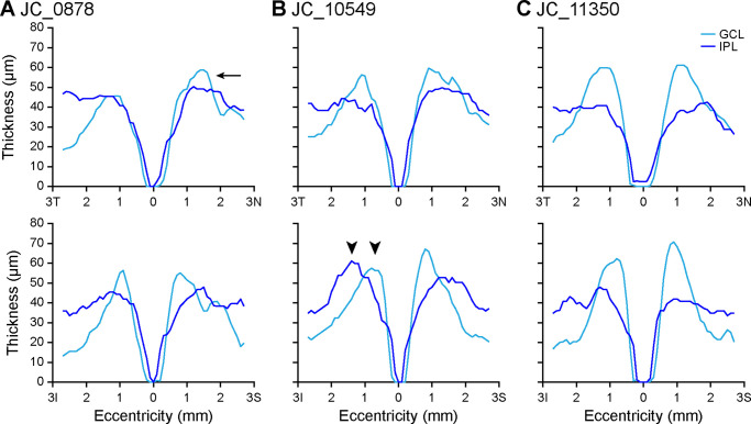
To test whether ganglion cell layer (GCL) and inner plexiform layer (IPL) topography is altered in albinism.

METHODS

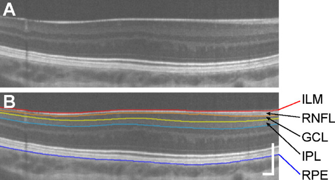
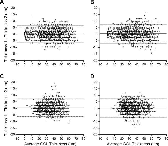
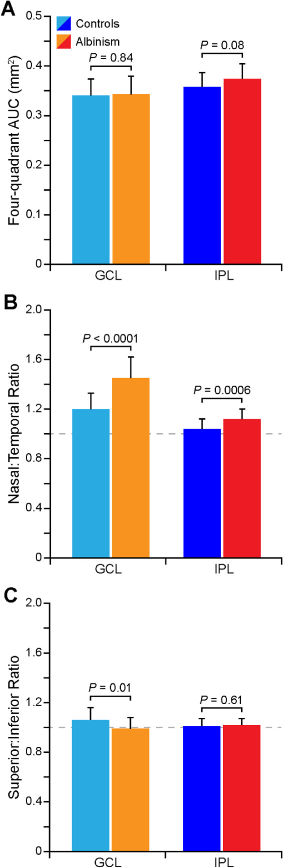
Optical coherence tomography scans were analyzed in 30 participants with albinism and 25 control participants. Horizontal and vertical line scans were acquired at the fovea, then strip registered and averaged. The Duke Optical Coherence Tomography Retinal Analysis Program was used to automatically segment the combined GCL and IPL and total retinal thickness, followed by program-assisted manual segmentation of the boundary between the GCL and IPL. Layer thickness and area under the curve (AUC) were calculated within 2.5 mm of the fovea. Nasal-temporal and superior-inferior asymmetry were calculated as an AUC ratio in each quadrant.

RESULTS

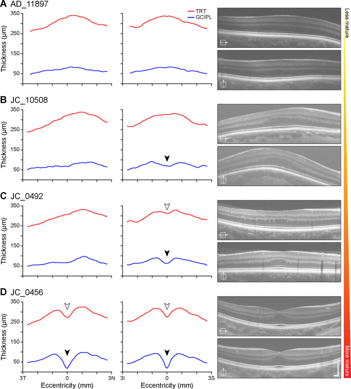
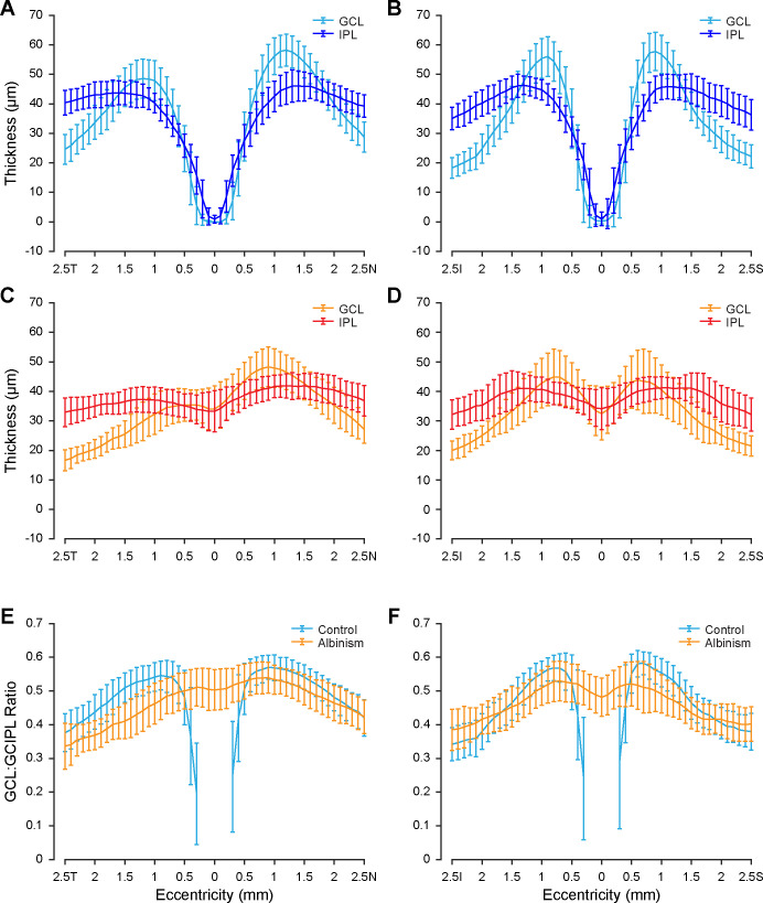
GCL and IPL topography varied between participants. The summed AUC in all quadrants was similar between groups for both the GCL (P = 0.84) and IPL (P = 0.08). Both groups showed nasal-temporal asymmetry in the GCL, but only participants with albinism had nasal-temporal asymmetry in the IPL. Nasal-temporal asymmetry was greater in albinism for both the GCL (P < 0.0001) and the IPL (P = 0.0006). The GCL usually comprised a greater percentage of the combined GCL and IPL in controls than in albinism.

CONCLUSIONS

The GCL and IPL have greater structural variability than previously reported. GCL and IPL topography are significantly altered in albinism, which suggests differences in the spatial distribution of retinal ganglion cells. This finding provides insight into foveal development and structure-function relationships in foveal hypoplasia.

摘要

目的

检测在白化病中神经节细胞层(GCL)和内丛状层(IPL)的地形是否发生改变。

方法

对 30 名白化病患者和 25 名对照者的光学相干断层扫描进行了分析。在黄斑区采集水平和垂直线扫描,然后进行条带配准和平均。使用 Duke 光学相干断层扫描视网膜分析程序自动分割 GCL 和 IPL 的总和以及总视网膜厚度,然后使用程序辅助手动分割 GCL 和 IPL 之间的边界。在距黄斑 2.5 毫米范围内计算层厚度和曲线下面积(AUC)。在每个象限中计算鼻颞侧和上-下侧的不对称性作为 AUC 比值。

结果

GCL 和 IPL 的地形在参与者之间存在差异。两组的 GCL(P = 0.84)和 IPL(P = 0.08)在所有象限的总和 AUC 相似。两组 GCL 均存在鼻颞侧不对称性,但只有白化病患者的 IPL 存在鼻颞侧不对称性。白化病患者的 GCL(P < 0.0001)和 IPL(P = 0.0006)的鼻颞侧不对称性更大。GCL 在对照组中通常占 GCL 和 IPL 总和的比例大于白化病患者。

结论

GCL 和 IPL 的结构变异性大于先前报道。GCL 和 IPL 在白化病中存在明显改变,这表明视网膜神经节细胞的空间分布存在差异。这一发现为黄斑发育和结构-功能关系提供了深入了解。

https://cdn.ncbi.nlm.nih.gov/pmc/blobs/6b6d/7405956/5f017f1c1208/iovs-61-3-36-f006.jpg
https://cdn.ncbi.nlm.nih.gov/pmc/blobs/6b6d/7405956/2c00b71a1bae/iovs-61-3-36-f001.jpg
https://cdn.ncbi.nlm.nih.gov/pmc/blobs/6b6d/7405956/32870796cd3c/iovs-61-3-36-f002.jpg
https://cdn.ncbi.nlm.nih.gov/pmc/blobs/6b6d/7405956/9e2c1a70d2ac/iovs-61-3-36-f003.jpg
https://cdn.ncbi.nlm.nih.gov/pmc/blobs/6b6d/7405956/3bdd219370f2/iovs-61-3-36-f004.jpg
https://cdn.ncbi.nlm.nih.gov/pmc/blobs/6b6d/7405956/a7e155f39a17/iovs-61-3-36-f005.jpg
https://cdn.ncbi.nlm.nih.gov/pmc/blobs/6b6d/7405956/5f017f1c1208/iovs-61-3-36-f006.jpg

相似文献

1
Assessing Ganglion Cell Layer Topography in Human Albinism Using Optical Coherence Tomography.利用光学相干断层扫描评估人类白化病的神经节细胞层形貌。
Invest Ophthalmol Vis Sci. 2020 Mar 9;61(3):36. doi: 10.1167/iovs.61.3.36.
2
Repeatability of peripapillary retinal nerve fiber layer and inner retinal thickness among two spectral domain optical coherence tomography devices.两种光谱域光学相干断层扫描设备测量视乳头周围视网膜神经纤维层及视网膜内层厚度的可重复性
Invest Ophthalmol Vis Sci. 2014 Sep 16;55(10):6536-46. doi: 10.1167/iovs.14-15072.
3
Structural evaluation of perimetrically normal and affected hemifields in open angle glaucoma.周边正常和青光眼受影响视野的结构评估。
Indian J Ophthalmol. 2019 Oct;67(10):1657-1662. doi: 10.4103/ijo.IJO_1755_18.
4
Ganglion cell layer-inner plexiform layer thickness and vision loss in young children with optic pathway gliomas.神经节细胞层-内丛状层厚度与视神经胶质瘤患儿视力丧失的关系。
Invest Ophthalmol Vis Sci. 2014 Mar 10;55(3):1402-8. doi: 10.1167/iovs.13-13119.
5
Ganglion Cell-Inner Plexiform Layer Thickness in Different Glaucoma Stages Measured by Optical Coherence Tomography.光学相干断层扫描测量不同青光眼阶段的神经节细胞-内网状层厚度
Ophthalmic Res. 2018;59(3):148-154. doi: 10.1159/000478052. Epub 2017 Sep 7.
6
Macular structure parameters as an automated indicator of paracentral scotoma in early glaucoma.黄斑结构参数作为早期青光眼旁中心暗点的自动指标。
Am J Ophthalmol. 2013 Nov;156(5):907-917.e1. doi: 10.1016/j.ajo.2013.06.029. Epub 2013 Aug 21.
7
Segmented inner plexiform layer thickness as a potential biomarker to evaluate open-angle glaucoma: Dendritic degeneration of retinal ganglion cell.分段内网状层厚度作为评估开角型青光眼的潜在生物标志物:视网膜神经节细胞的树突状变性
PLoS One. 2017 Aug 3;12(8):e0182404. doi: 10.1371/journal.pone.0182404. eCollection 2017.
8
Distribution of macular ganglion cell layer thickness in foveal hypoplasia: A new diagnostic criterion for ocular albinism.黄斑神经节细胞层厚度在视盘发育不良中的分布:一种新的眼白化病诊断标准。
PLoS One. 2019 Nov 18;14(11):e0224410. doi: 10.1371/journal.pone.0224410. eCollection 2019.
9
Retinal Ganglion Cell Topography in Patients With Visual Pathway Pathology.视路病变患者的视网膜神经节细胞层地形图。
J Neuroophthalmol. 2018 Jun;38(2):172-178. doi: 10.1097/WNO.0000000000000589.
10
Comparative analysis of mean retinal thickness measured using SD-OCT in normal young or old age and glaucomatous eyes.使用频域光学相干断层扫描(SD-OCT)测量的正常年轻人或老年人以及青光眼患者眼睛的平均视网膜厚度的比较分析。
Int Ophthalmol. 2018 Dec;38(6):2417-2426. doi: 10.1007/s10792-017-0744-7. Epub 2017 Oct 12.

引用本文的文献

1
Quantitative Foveal Structural Metrics as Predictors of Visual Acuity in Human Albinism.定量黄斑结构测量指标可预测人类白化病的视力。
Invest Ophthalmol Vis Sci. 2024 Mar 5;65(3):3. doi: 10.1167/iovs.65.3.3.
2
Temporal-to-Nasal Macular Ganglion Cell and Inner Plexiform Layer Ratios in a Large Adult Twin Cohort: Correlations With Age and Heritability.大样本成人双胞胎队列中颞侧至鼻侧黄斑神经节细胞和内丛状层的比率:与年龄和遗传度的相关性。
Invest Ophthalmol Vis Sci. 2024 Feb 1;65(2):26. doi: 10.1167/iovs.65.2.26.
3
Visual Field Deficits in Albinism in Comparison to Idiopathic Infantile Nystagmus.

本文引用的文献

1
Distribution of macular ganglion cell layer thickness in foveal hypoplasia: A new diagnostic criterion for ocular albinism.黄斑神经节细胞层厚度在视盘发育不良中的分布:一种新的眼白化病诊断标准。
PLoS One. 2019 Nov 18;14(11):e0224410. doi: 10.1371/journal.pone.0224410. eCollection 2019.
2
Improving visible light OCT of the human retina with rapid spectral shaping and axial tracking.通过快速光谱整形和轴向跟踪改善人眼视网膜的可见光光学相干断层扫描
Biomed Opt Express. 2019 May 21;10(6):2918-2931. doi: 10.1364/BOE.10.002918. eCollection 2019 Jun 1.
3
Aberrant visual pathway development in albinism: From retina to cortex.
白化病与特发性婴儿眼球震颤的视野缺损比较
Invest Ophthalmol Vis Sci. 2024 Feb 1;65(2):13. doi: 10.1167/iovs.65.2.13.
4
Phenotypic Features Determining Visual Acuity in Albinism and the Role of Amblyogenic Factors.白化病中决定视力的表型特征及致弱视因素的作用
Invest Ophthalmol Vis Sci. 2024 Feb 1;65(2):14. doi: 10.1167/iovs.65.2.14.
5
Clinical features and imaging characteristics in achiasmia.失认症的临床特征与影像学特点
Brain Commun. 2023 Aug 22;5(4):fcad219. doi: 10.1093/braincomms/fcad219. eCollection 2023.
6
Optical Coherence Tomography as a Biomarker for Differential Diagnostics in Nystagmus: Ganglion Cell Layer Thickness Ratio.光学相干断层扫描作为眼球震颤鉴别诊断的生物标志物:神经节细胞层厚度比
J Clin Med. 2022 Aug 23;11(17):4941. doi: 10.3390/jcm11174941.
7
Retinal alterations in patients with Lafora disease.拉福拉病患者的视网膜改变。
Am J Ophthalmol Case Rep. 2021 Jun 15;23:101146. doi: 10.1016/j.ajoc.2021.101146. eCollection 2021 Sep.
8
Examining Whether AOSLO-Based Foveal Cone Metrics in Achromatopsia and Albinism Are Representative of Foveal Cone Structure.检查基于 AOSLO 的先天性静止性夜盲症和白化病的黄斑中心凹锥细胞测量指标是否代表黄斑中心凹锥细胞结构。
Transl Vis Sci Technol. 2021 May 3;10(6):22. doi: 10.1167/tvst.10.6.22.
9
Morphometry of Inner Plexiform Layer (IPL) Stratification in the Human Retina With Visible Light Optical Coherence Tomography.利用可见光光学相干断层扫描技术对人视网膜内网状层(IPL)分层进行形态测量
Front Cell Neurosci. 2021 Apr 29;15:655096. doi: 10.3389/fncel.2021.655096. eCollection 2021.
10
Characterization of Retinal Thickness in Individuals with Albinism: Baseline Data for a Black South African Population.白化病患者视网膜厚度的特征:南非黑人人群的基线数据。
Clin Optom (Auckl). 2021 Jan 20;13:15-22. doi: 10.2147/OPTO.S273141. eCollection 2021.
白化病中异常的视觉通路发育:从视网膜到皮层。
Hum Brain Mapp. 2019 Feb 15;40(3):777-788. doi: 10.1002/hbm.24411. Epub 2018 Dec 4.
4
Macular ganglion cell asymmetry for detecting paracentral scotoma in early glaucoma.用于检测早期青光眼旁中心暗点的黄斑神经节细胞不对称性。
Clin Ophthalmol. 2018 Nov 5;12:2253-2260. doi: 10.2147/OPTH.S181551. eCollection 2018.
5
The Henle Fiber Layer in Albinism: Comparison to Normal and Relationship to Outer Nuclear Layer Thickness and Foveal Cone Density.白化病中的 Henle 纤维层:与正常的比较以及与外核层厚度和中心凹锥密度的关系。
Invest Ophthalmol Vis Sci. 2018 Nov 1;59(13):5336-5348. doi: 10.1167/iovs.18-24145.
6
The Fovea-BMO Axis Angle and Macular Thickness Vertical Asymmetry Across The Temporal Raphe.黄斑中心凹-BMO 轴夹角与沿 temporal raphe 的黄斑厚度垂直不对称性。
J Glaucoma. 2018 Nov;27(11):993-998. doi: 10.1097/IJG.0000000000001077.
7
The Phenotypic Spectrum of Albinism.白化病表型谱。
Ophthalmology. 2018 Dec;125(12):1953-1960. doi: 10.1016/j.ophtha.2018.08.003. Epub 2018 Aug 8.
8
Ultrahigh resolution retinal imaging by visible light OCT with longitudinal achromatization.通过具有纵向消色差功能的可见光光学相干断层扫描实现超高分辨率视网膜成像。
Biomed Opt Express. 2018 Mar 5;9(4):1477-1491. doi: 10.1364/BOE.9.001477. eCollection 2018 Apr 1.
9
Imaging and quantifying ganglion cells and other transparent neurons in the living human retina.在活体人视网膜中成像和定量神经节细胞和其他透明神经元。
Proc Natl Acad Sci U S A. 2017 Nov 28;114(48):12803-12808. doi: 10.1073/pnas.1711734114. Epub 2017 Nov 14.
10
Macular imaging by optical coherence tomography in the diagnosis and management of glaucoma.光学相干断层扫描在青光眼诊断和管理中的黄斑成像。
Br J Ophthalmol. 2018 Jun;102(6):718-724. doi: 10.1136/bjophthalmol-2017-310869. Epub 2017 Oct 21.