Hou Yonghui, Lin Weiping, Li Ying, Sun Yuxin, Liu Yamei, Chen Chen, Jiang Xiaohua, Li Gang, Xu Liangliang
Key Laboratory of Orthopaedics & Traumatology, Guangdong Provincial Hospital of Chinese Medicine, The Second Affiliated Hospital of Guangzhou University of Chinese Medicine, Guangzhou, China.
Lingnan Medical Research Center, The First Affiliated Hospital of Guangzhou University of Chinese Medicine, Guangzhou University of Chinese Medicine, Guangzhou, China.
J Orthop Translat. 2020 Dec 10;27:25-32. doi: 10.1016/j.jot.2020.10.009. eCollection 2021 Mar.
Mesenchymal stem cells (MSCs) are promising targets for therapeutic use in regenerative medicine and tissue engineering. In the previous study, we have found that MSCs could be reverted to a primitive stem cell population after in vitro induction of osteogenic and de-osteogenic differentiation (de-osteogenic differentiated MSCs, De-Os-MSCs). De-Os-MSCs showed improved cell survival and osteogenic potential. However, the underlying mechanism and its potential effect on fracture healing has not been explored.
MSCs were isolated from the rat bone marrow. MicroRNAs were cloned into lentiviral vectors and transduced into MSCs to observe the effects on osteogenesis. The expression levels of marker genes were evaluated by quantitative RT-PCR. Ectopic bone formation model was used to evaluate the bone regeneration ability of mir-92b transduced MSCs in vivo. An open femur fracture model was established, and MSCs or De-Os-MSCs were administrated to the fracture sites. Histological, biomechanical and microCT analysis were used to evaluate the quality of bone.
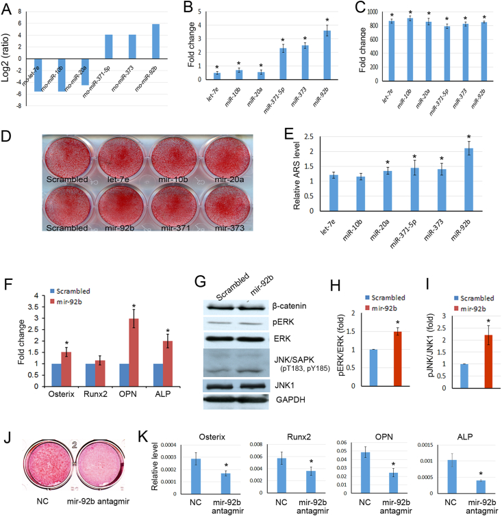
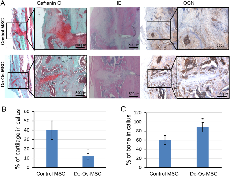
In the present study, we found that mir-92b was significantly increased in the secretions of De-Os-MSCs. And mir-92b could promote the osteogenic differentiation potential of MSCs by activating pERK and JNK signaling pathways. The ectopic bone formation assay showed that MSCs overexpressing mir-92b formed more bone like tissues in vivo. Most importantly, we found local administration of De-Os-MSCs could accelerate fracture healing using an open femur fracture model in rats. The quality of bone property was much better as shown by microCT and biomechanical testing.
Taken together, our study demonstrated that mir-92b promoted osteogenesis of MSCs, which was partially accounted for the enhanced osteogenic differentiation potential of De-Os-MSCs. And De-Os-MSCs had shown better regenerative capacity in accelerating fracture healing when they were locally given.
De-Os-MSCs could be used to accelerate fracture healing, and reduce the occurrence of delayed unions and non-unions.
间充质干细胞(MSCs)是再生医学和组织工程中有前景的治疗靶点。在先前的研究中,我们发现间充质干细胞在体外诱导成骨和去成骨分化后(去成骨分化的间充质干细胞,De-Os-MSCs)可恢复为原始干细胞群体。De-Os-MSCs表现出更好的细胞存活能力和成骨潜能。然而,其潜在机制及其对骨折愈合的潜在影响尚未得到探索。
从大鼠骨髓中分离间充质干细胞。将微小RNA克隆到慢病毒载体中并转导至间充质干细胞中,以观察其对成骨的影响。通过定量逆转录聚合酶链反应评估标记基因的表达水平。采用异位骨形成模型评估转导mir-92b的间充质干细胞在体内的骨再生能力。建立开放性股骨骨折模型,并将间充质干细胞或De-Os-MSCs应用于骨折部位。采用组织学、生物力学和显微CT分析评估骨质量。
在本研究中,我们发现mir-92b在De-Os-MSCs的分泌物中显著增加。并且mir-92b可通过激活pERK和JNK信号通路促进间充质干细胞的成骨分化潜能。异位骨形成试验表明,过表达mir-92b的间充质干细胞在体内形成更多类骨组织。最重要的是,我们发现使用大鼠开放性股骨骨折模型局部应用De-Os-MSCs可加速骨折愈合。显微CT和生物力学测试显示骨特性质量更好。
综上所述,我们的研究表明mir-92b促进间充质干细胞的成骨作用,这部分解释了De-Os-MSCs增强的成骨分化潜能。并且当局部给予De-Os-MSCs时,其在加速骨折愈合方面表现出更好的再生能力。
De-Os-MSCs可用于加速骨折愈合,并减少延迟愈合和不愈合的发生。