Guzzetti Ezequiel, Annabi Mohamed-Salah, Pibarot Philippe, Clavel Marie-Annick
Institut Universitaire de Cardiologie et de Pneumologie de Québec (Quebec Heart & Lung Institute), Quebec, QC, Canada.
Front Cardiovasc Med. 2020 Dec 3;7:570689. doi: 10.3389/fcvm.2020.570689. eCollection 2020.
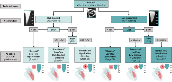
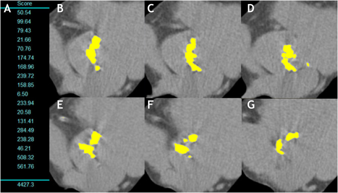
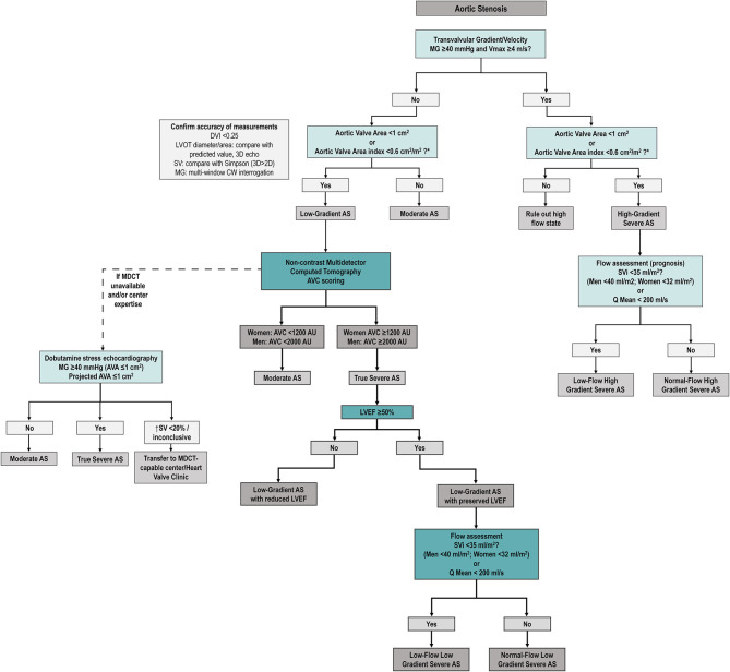
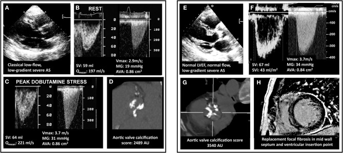
Aortic stenosis (AS) is a disease of the valve and the myocardium. A correct assessment of the valve disease severity is key to define the need for aortic valve replacement (AVR), but a better understanding of the myocardial consequences of the increased afterload is paramount to optimize the timing of the intervention. Transthoracic echocardiography remains the cornerstone of AS assessment, as it is universally available, and it allows a comprehensive structural and hemodynamic evaluation of both the aortic valve and the rest of the heart. However, it may not be sufficient as a significant proportion of patients with severe AS presents with discordant grading (i.e., an AVA ≤ 1 cm and a mean gradient <40 mmHg) which raises uncertainty about the true severity of AS and the need for AVR. Several imaging modalities (transesophageal or stress echocardiography, computed tomography, cardiovascular magnetic resonance, positron emission tomography) exist that allow a detailed assessment of the stenotic aortic valve and the myocardial remodeling response. This review aims to provide an updated overview of these multimodality imaging techniques and seeks to highlight a practical approach to help clinical decision making in the challenging group of patients with discordant low-gradient AS.
主动脉瓣狭窄(AS)是一种累及瓣膜和心肌的疾病。准确评估瓣膜疾病的严重程度是确定是否需要进行主动脉瓣置换术(AVR)的关键,但更好地了解后负荷增加对心肌的影响对于优化干预时机至关重要。经胸超声心动图仍然是AS评估的基石,因为它普遍可用,并且能够对主动脉瓣和心脏其他部分进行全面的结构和血流动力学评估。然而,它可能并不足够,因为相当一部分重度AS患者存在分级不一致的情况(即主动脉瓣口面积≤1 cm且平均压差<40 mmHg),这增加了对AS真正严重程度以及AVR必要性的不确定性。现有的几种成像方式(经食管或负荷超声心动图、计算机断层扫描、心血管磁共振、正电子发射断层扫描)能够对狭窄的主动脉瓣和心肌重塑反应进行详细评估。本综述旨在提供这些多模态成像技术的最新概述,并试图突出一种实用方法实用方法,以帮助在具有分级不一致的低压差AS这一具有挑战性的患者群体中进行临床决策。