University of Bordeaux, Centre de Recherche Cardio-Thoracique de Bordeaux, U1045, CIC 1401, Bordeaux, France; INSERM, Centre de Recherche Cardio-Thoracique de Bordeaux, U1045, CIC 1401, Bordeaux, France; CHU de Bordeaux, Service d'Imagerie Thoracique et Cardiovasculaire, Service des Maladies Respiratoires, Service d'Exploration Fonctionnelle Respiratoire, CIC 1401, Pessac, France; Center for Pulmonary Imaging Research, Division of Pulmonary Medicine and Department of Radiology, Cincinnati Children's Hospital Medical Center, Cincinnati, OH.
Center for Pulmonary Imaging Research, Division of Pulmonary Medicine and Department of Radiology, Cincinnati Children's Hospital Medical Center, Cincinnati, OH; Department of Pediatrics, College of Medicine, University of Cincinnati, Cincinnati, OH.
Chest. 2021 Jun;159(6):2205-2217. doi: 10.1016/j.chest.2020.12.008. Epub 2020 Dec 17.
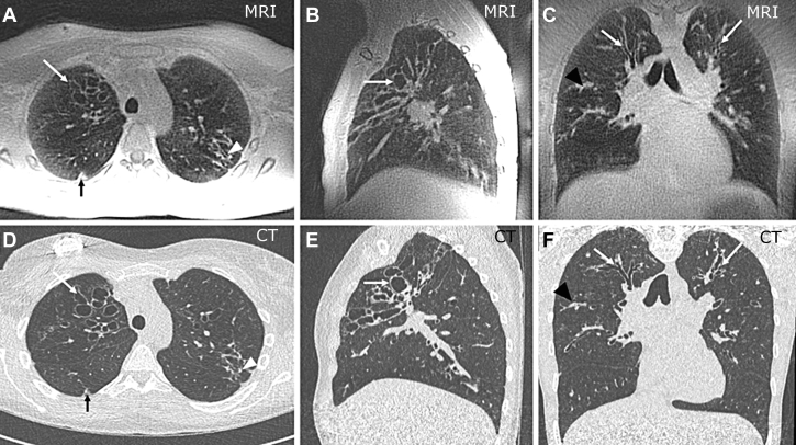
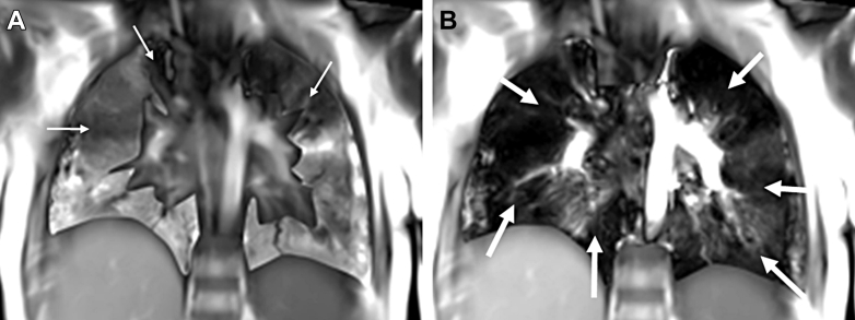
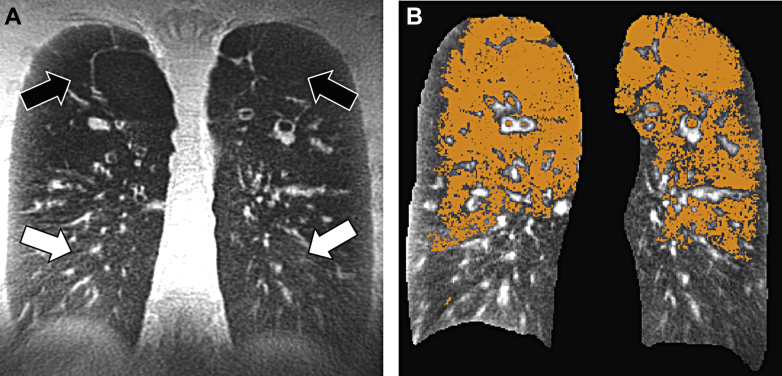
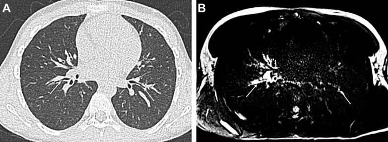
To assess airway and lung parenchymal damage noninvasively in cystic fibrosis (CF), chest MRI has been historically out of the scope of routine clinical imaging because of technical difficulties such as low proton density and respiratory and cardiac motion. However, technological breakthroughs have emerged that dramatically improve lung MRI quality (including signal-to-noise ratio, resolution, speed, and contrast). At the same time, novel treatments have changed the landscape of CF clinical care. In this contemporary context, there is now consensus that lung MRI can be used clinically to assess CF in a radiation-free manner and to enable quantification of lung disease severity. MRI can now achieve three-dimensional, high-resolution morphologic imaging, and beyond this morphologic information, MRI may offer the ability to sensitively differentiate active inflammation vs scarring tissue. MRI could also characterize various forms of inflammation for early guidance of treatment. Moreover, functional information from MRI can be used to assess regional, small-airway disease with sensitivity to detect small changes even in patients with mild CF. Finally, automated quantification methods have emerged to support conventional visual analyses for more objective and reproducible assessment of disease severity. This article aims to review the most recent developments of lung MRI, with a focus on practical application and clinical value in CF, and the perspectives on how these modern techniques may converge and impact patient care soon.
为了在囊性纤维化 (CF) 中无创地评估气道和肺实质损伤,胸部 MRI 由于质子密度低以及呼吸和心脏运动等技术难题,历史上一直超出常规临床成像的范围。然而,技术突破已经出现,极大地提高了肺部 MRI 的质量(包括信噪比、分辨率、速度和对比度)。与此同时,新型治疗方法改变了 CF 临床护理的格局。在这个现代背景下,现在已经达成共识,即肺 MRI 可以在临床中以无辐射的方式用于评估 CF,并能够定量评估肺部疾病的严重程度。MRI 现在可以实现三维、高分辨率的形态成像,并且超越这种形态信息,MRI 可能具有区分活跃炎症与瘢痕组织的能力。MRI 还可以对各种形式的炎症进行特征描述,以便尽早指导治疗。此外,MRI 的功能信息可用于评估区域性、小气道疾病,其敏感性足以检测到即使是轻度 CF 患者的微小变化。最后,已经出现了自动化定量方法,以支持常规视觉分析,从而更客观、更可重复地评估疾病严重程度。本文旨在综述肺部 MRI 的最新进展,重点介绍其在 CF 中的实际应用和临床价值,以及这些现代技术如何融合并对患者护理产生影响的前景。