Ostrycharz Ewa, Wasik Urszula, Kempinska-Podhorodecka Agnieszka, Banales Jesus M, Milkiewicz Piotr, Milkiewicz Malgorzata
Department of Medical Biology, Pomeranian Medical University, 71-111 Szczecin, Poland.
Department of Liver and Gastrointestinal Diseases, Biodonostia Health Research Institute-Donostia University Hospital-Ikerbasque, CIBERehd, University of the Basque Country (UPV/EHU), 20014 San Sebastian, Spain.
Int J Mol Sci. 2020 Dec 18;21(24):9667. doi: 10.3390/ijms21249667.
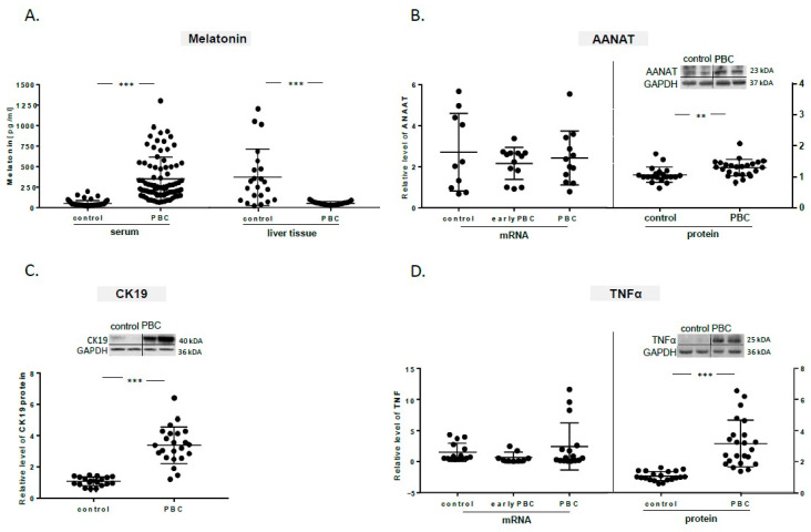
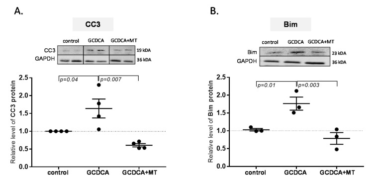
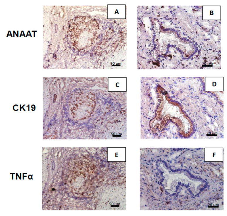
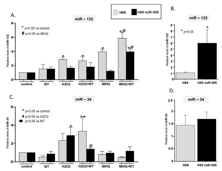
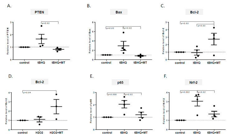
Biosynthesis of melatonin by cholangiocytes is essential for maintaining the function of biliary epithelium. However, this cytoprotective mechanism appears to be impaired in primary biliary cholangitis (PBC). MiR-132 has emerged as a mediator of inflammation in chronic liver diseases. The effect of melatonin on oxidative stress and bile acid-induced apoptosis was also examined in cholangiocyes overexpressing miR506, as a PBC-like cellular model. In PBC patients the serum levels of melatonin were found increased in comparison to healthy controls. Whereas, in cholangiocytes within cirrhotic PBC livers the melatonin biosynthetic pathway was substantially suppressed even though the expressions of melatonin rate-limiting enzyme aralkylamine N-acetyltransferase (AANAT), and CK-19 (marker of cholangiocytes) were enhanced. In cholangiocytes exposed to mitochondrial oxidative stress melatonin decreased the expression of proapoptotic stimuli (PTEN, Bax, miR-34), which was accompanied by the inhibition of a pivotal mediator of inflammatory response Nf-κB-p65 and the activation of antiapoptotic signaling (miR-132, Bcl2). Similarly, melatonin reduced bile acid-induced proapoptotic caspase 3 and Bim levels. In summary, the insufficient hepatic expression of melatonin in PBC patients may predispose cholangiocytes to oxidative stress-related damage. Melatonin, via epigenetic modulation, was able to suppress NF-κB signaling activation and protect against biliary cells apoptotic signaling.
胆管细胞合成褪黑素对于维持胆管上皮功能至关重要。然而,这种细胞保护机制在原发性胆汁性胆管炎(PBC)中似乎受损。MiR-132已成为慢性肝病炎症的介质。作为PBC样细胞模型,还在过表达miR506的胆管细胞中研究了褪黑素对氧化应激和胆汁酸诱导的细胞凋亡的影响。在PBC患者中,发现血清褪黑素水平比健康对照者升高。然而,在肝硬化PBC肝脏中的胆管细胞中,尽管褪黑素限速酶芳烷基胺N-乙酰转移酶(AANAT)和CK-19(胆管细胞标志物)的表达增强,但褪黑素生物合成途径却被大幅抑制。在暴露于线粒体氧化应激的胆管细胞中,褪黑素降低了促凋亡刺激物(PTEN、Bax、miR-34)的表达,同时伴随着炎症反应关键介质Nf-κB-p65的抑制和抗凋亡信号(miR-132、Bcl2)的激活。同样,褪黑素降低了胆汁酸诱导促凋亡的半胱天冬酶3和Bim水平。总之,PBC患者肝脏中褪黑素表达不足可能使胆管细胞易受氧化应激相关损伤。褪黑素通过表观遗传调控,能够抑制NF-κB信号激活并保护胆管细胞免受凋亡信号影响。