Section of Rehabilitation, Kanazawa University Hospital, Ishikawa, Japan.
Department of Motor Function Analysis, Human Health Sciences, Graduate School of Medicine, Kyoto University, Kyoto, Japan.
Cartilage. 2021 Dec;13(2_suppl):1522S-1529S. doi: 10.1177/1947603520982350. Epub 2020 Dec 27.
The study aim was to evaluate the histological relationship between osteoarthritis (OA) and articular cartilage in disuse atrophy induced by hindlimb unloading in a post-traumatic OA rat model.
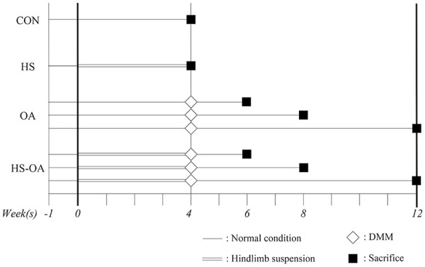
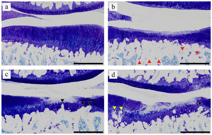
Forty male rats were divided into the 4 following experimental groups: control, hindlimb suspension (HS), OA induced by destabilization of the medial meniscus (OA), and OA induction after hindlimb suspension (HS-OA). Histological changes in the articular cartilage of the tibia were evaluated by the Osteoarthritis Research Society International (OARSI) scores and histomorphometrical analyses at 2, 4, and 8 weeks after OA induction.
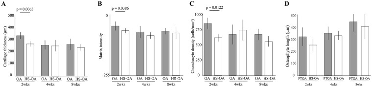
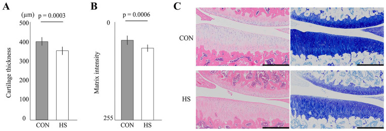
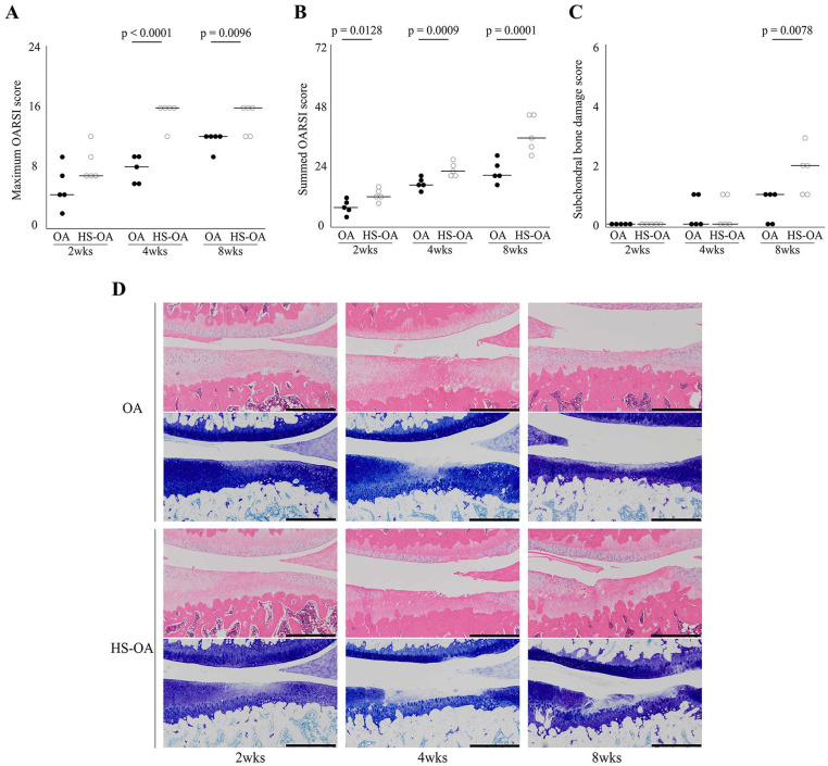
We confirmed that disuse atrophy of the articular cartilage was caused by thinning of the articular cartilage and the decrease in matrix staining for the nonloading period of 4 weeks. The OARSI scores and histomorphological analyses revealed that OA progressed significantly wider and deeper in the HS-OA group than in the OA group over time. In the sham group, disuse atrophy of the articular cartilage recovered at 2 weeks after reloading.
This study revealed that OA progressed faster in cartilage atrophy than in normal articular cartilage. Further studies are required for investigating the mechanisms of disuse atrophy of cartilage and its association with OA using the biochemical and immunohistochemical analysis.
本研究旨在评估在创伤后骨关节炎大鼠模型中,由后肢去负荷引起的废用性萎缩对骨关节炎(OA)和关节软骨的组织学关系。
40 只雄性大鼠分为以下 4 个实验组:对照组、后肢悬吊(HS)组、内侧半月板不稳定诱导的 OA(OA)组和后肢悬吊后诱导的 OA(HS-OA)组。在 OA 诱导后 2、4 和 8 周,通过 OARSI 评分和组织形态计量学分析评估胫骨关节软骨的组织学变化。
我们证实,关节软骨的废用性萎缩是由非负荷期 4 周内软骨变薄和基质染色减少引起的。OARSI 评分和组织形态学分析显示,随着时间的推移,HS-OA 组的 OA 进展明显比 OA 组更广泛和更深。在假手术组中,在重新加载后 2 周,关节软骨的废用性萎缩得到恢复。
本研究表明,OA 在软骨萎缩中的进展比正常关节软骨更快。需要进一步研究使用生化和免疫组织化学分析来研究软骨废用性萎缩及其与 OA 的关系的机制。