You Sungyong, Chao Jerry, Cohen Edward A K, Ward E Sally, Ober Raimund J
Opt Express. 2021 Jan 4;29(1):182-207. doi: 10.1364/OE.408361.
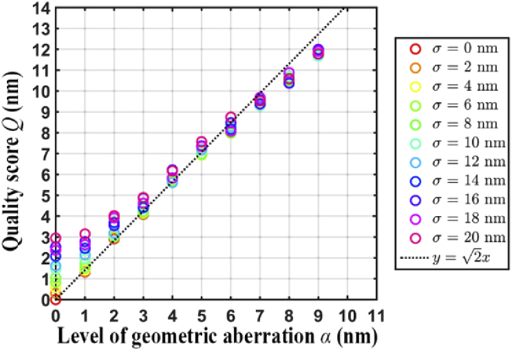
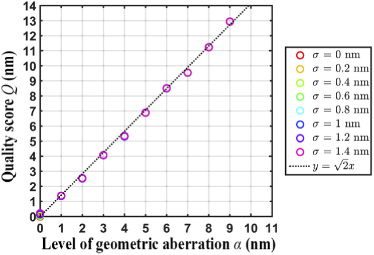
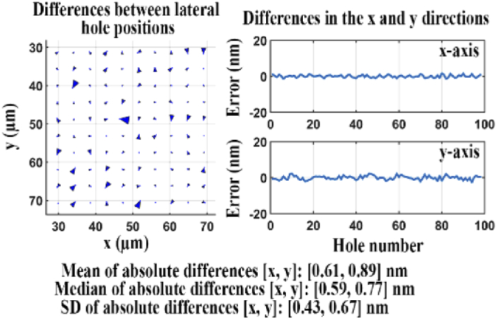
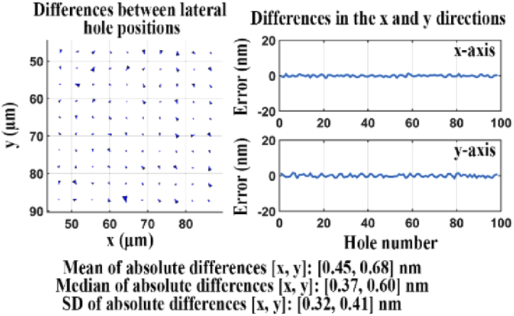
Single-molecule microscopy allows for the investigation of the dynamics of individual molecules and the visualization of subcellular structures at high spatial resolution. For single-molecule imaging experiments, and particularly those that entail the acquisition of multicolor data, calibration of the microscope and its optical components therefore needs to be carried out at a high level of accuracy. We propose here a method for calibrating a microscope at the nanometer scale, in the sense of determining optical aberrations as revealed by point source localization errors on the order of nanometers. The method is based on the imaging of a standard sample to detect and evaluate the amount of geometric aberration introduced in the optical light path. To provide support for multicolor imaging, it also includes procedures for evaluating the geometric aberration caused by a dichroic filter and the axial chromatic aberration introduced by an objective lens.
单分子显微镜能够研究单个分子的动力学,并以高空间分辨率可视化亚细胞结构。因此,对于单分子成像实验,尤其是那些需要采集多色数据的实验,显微镜及其光学组件的校准需要高精度进行。我们在此提出一种在纳米尺度上校准显微镜的方法,即确定由纳米级点源定位误差所揭示的光学像差。该方法基于对标准样品的成像,以检测和评估在光学光路中引入的几何像差量。为支持多色成像,它还包括评估二向色滤光片引起的几何像差以及物镜引入的轴向色差的程序。