Vannini Eleonora, Restani Laura, Dilillo Marialaura, McDonnell Liam A, Caleo Matteo, Marra Vincenzo
Neuroscience Institute, National Research Council (CNR), Pisa, Italy.
Department of Neuroscience, Psychology and Behaviour, University of Leicester, Leicester, United Kingdom.
Front Cell Neurosci. 2020 Dec 10;14:606142. doi: 10.3389/fncel.2020.606142. eCollection 2020.
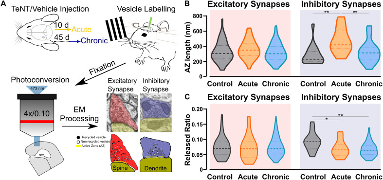
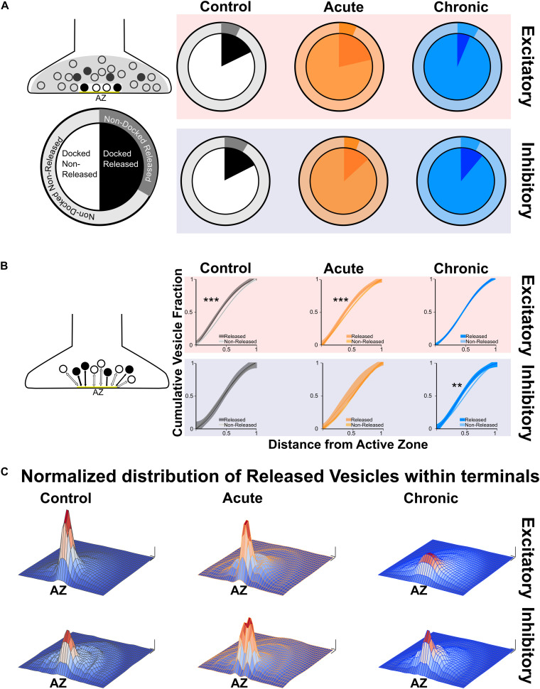
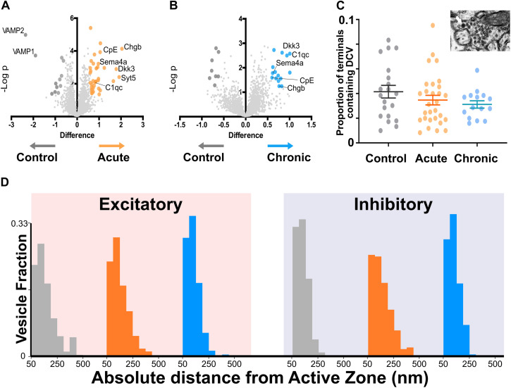
Neuronal hyperexcitability often results from an unbalance between excitatory and inhibitory neurotransmission, but the synaptic alterations leading to enhanced seizure propensity are only partly understood. Taking advantage of a mouse model of neocortical epilepsy, we used a combination of photoconversion and electron microscopy to assess changes in synaptic vesicles pools . Our analyses reveal that epileptic networks show an early onset lengthening of active zones at inhibitory synapses, together with a delayed spatial reorganization of recycled vesicles at excitatory synapses. Proteomics of synaptic content indicate that specific proteins were increased in epileptic mice. Altogether, our data reveal a complex landscape of nanoscale changes affecting the epileptic synaptic release machinery. In particular, our findings show that an altered positioning of release-competent vesicles represent a novel signature of epileptic networks.
神经元的过度兴奋通常源于兴奋性和抑制性神经传递之间的失衡,但导致癫痫易感性增强的突触改变仅得到部分理解。利用一种新皮质癫痫小鼠模型,我们结合光转换和电子显微镜来评估突触小泡池的变化。我们的分析表明,癫痫网络在抑制性突触处显示出活性区早期开始延长,同时在兴奋性突触处回收小泡的空间重组延迟。突触内容物的蛋白质组学表明,癫痫小鼠中特定蛋白质增加。总之,我们的数据揭示了影响癫痫突触释放机制的纳米级变化的复杂情况。特别是,我们的研究结果表明,具有释放能力的小泡定位改变是癫痫网络的一种新特征。