Yun Taesik, Lee Kang-Il, Koo Yoonhoi, Kim Hakhyun, Chang Dongwoo, Lee Chulhyun, Yang Mhan-Pyo, Kang Byeong-Teck
Laboratory of Veterinary Internal Medicine, College of Veterinary Medicine, Chungbuk National University, Cheongju, South Korea.
Section of Veterinary Medical Imaging, Veterinary Teaching Hospital, College of Veterinary Medicine, Chungbuk National University, Cheongju, South Korea.
Front Vet Sci. 2020 Dec 10;7:598792. doi: 10.3389/fvets.2020.598792. eCollection 2020.
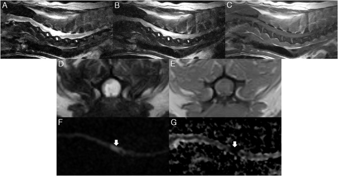
A 9-year-old, intact male Shih Tzu dog presented with systemic weakness and peracute onset of tetraplegia. Tetraplegia with lower motor neuron signs was noted upon neurological examination. Diseases that cause acute flaccid tetraparesis, such as acute fulminating myasthenia gravis, polyradiculoneuritis, tick paralysis, and botulism, were ruled out based on the medical history, normal electrophysiological tests, and non-response to the neostigmine challenging test. Initial 0.3-Tesla (T) magnetic resonance imaging (MRI) findings included sharply demarcated intramedullary lesions at the C3-C6 level, mainly involving gray matter, which appeared hypo- to iso- intense on T1-weighted images (WIs), and hyperintense on T2-WIs and fluid-attenuated inversion recovery images. There was no enhancement on post-contrast T1-WIs. Neutrophilic pleocytosis was observed in the cerebrospinal fluid analysis. No clinical responses were observed for the treatment of non-infectious myelitis with an immunosuppressive dosage of prednisolone. A follow-up 3-T MRI 6 days later demonstrated hyperintensity on diffusion-WI (DWI) and a decreased apparent diffusion coefficient (ADC) value (0.54 × 10 mm/s) of the spinal lesions. Through histological examination, a fibrocartilaginous embolism was definitively confirmed. This is the first report to describe an ischemic spinal injury visualized by DWI and ADC mapping with high-field MRI in a chondrodystrophic dog diagnosed with a fibrocartilaginous embolism.
一只9岁未绝育的雄性西施犬出现全身无力和急性四肢瘫痪。神经学检查发现四肢瘫痪并伴有下运动神经元体征。根据病史、正常的电生理检查结果以及对新斯的明激发试验无反应,排除了导致急性弛缓性四肢轻瘫的疾病,如急性暴发性重症肌无力、多神经根神经炎、蜱瘫痪和肉毒中毒。最初的0.3特斯拉(T)磁共振成像(MRI)结果显示,在C3 - C6水平有界限清晰的髓内病变,主要累及灰质,在T1加权像(WI)上呈低到等信号,在T2加权像和液体衰减反转恢复图像上呈高信号。增强后的T1加权像上无强化。脑脊液分析显示中性粒细胞增多。使用免疫抑制剂量的泼尼松龙治疗非感染性脊髓炎未见临床反应。6天后的3T MRI随访显示,脊髓病变在扩散加权像(DWI)上呈高信号,表观扩散系数(ADC)值降低(0.54×10⁻³mm²/s)。通过组织学检查,最终确诊为纤维软骨栓塞。这是第一份描述在一只被诊断为纤维软骨栓塞的软骨发育不良犬中,通过高场MRI的DWI和ADC映射观察到缺血性脊髓损伤的报告。