CONICET, Instituto de Química Biológica de la Facultad de Ciencias Exactas y Naturales (IQUIBICEN), Universidad de Buenos Aires, Buenos Aires C1428EGA, Argentina.
Departamento de Química Biológica, Facultad de Ciencias Exactas y Naturales, Universidad de Buenos Aires, Buenos Aires C1428EGA, Argentina.
Cells. 2020 Dec 29;10(1):35. doi: 10.3390/cells10010035.
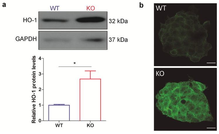
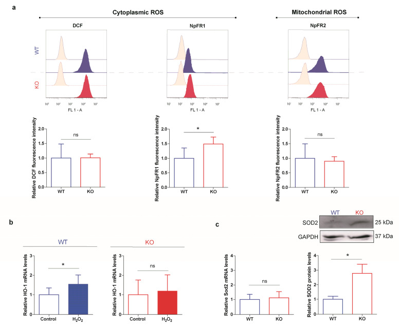
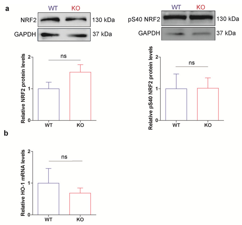
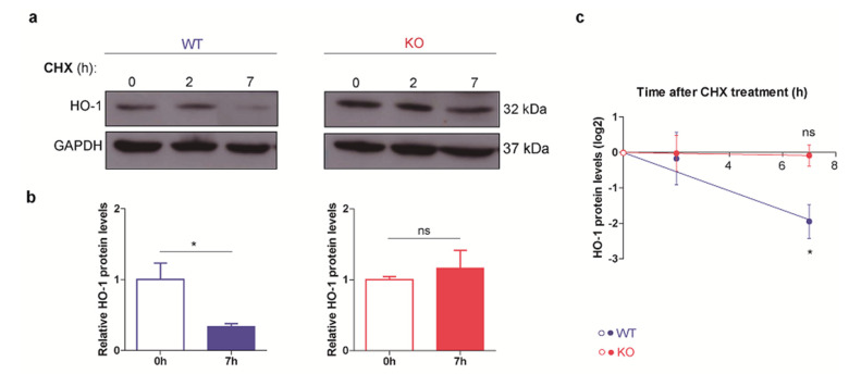
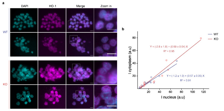
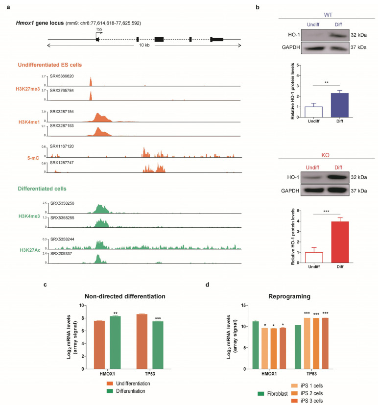
Stem cells genome safeguarding requires strict oxidative stress control. Heme oxygenase-1 (HO-1) and p53 are relevant components of the cellular defense system. p53 controls cellular response to multiple types of harmful stimulus, including oxidative stress. Otherwise, besides having a protective role, HO-1 is also involved in embryo development and in embryonic stem (ES) cells differentiation. Although both proteins have been extensively studied, little is known about their relationship in stem cells. The aim of this work is to explore HO-1-p53 interplay in ES cells. We studied HO-1 expression in p53 knockout (KO) ES cells and we found that they have higher HO-1 protein levels but similar HO-1 mRNA levels than the wild type (WT) ES cell line. Furthermore, cycloheximide treatment increased HO-1 abundance in p53 KO cells suggesting that p53 modulates HO-1 protein stability. Notably, HO treatment did not induce HO-1 expression in p53 KO ES cells. Finally, SOD2 protein levels are also increased while transcripts are not in KO cells, further suggesting that the p53 null phenotype is associated with a reinforcement of the antioxidant machinery. Our results demonstrate the existence of a connection between p53 and HO-1 in ES cells, highlighting the relationship between these stress defense pathways.
干细胞基因组的保护需要严格控制氧化应激。血红素加氧酶-1(HO-1)和 p53 是细胞防御系统的相关组成部分。p53 控制细胞对多种有害刺激(包括氧化应激)的反应。此外,HO-1 除了具有保护作用外,还参与胚胎发育和胚胎干细胞(ES)细胞分化。尽管这两种蛋白质已经被广泛研究,但它们在干细胞中的关系知之甚少。本研究旨在探讨 ES 细胞中 HO-1-p53 的相互作用。我们研究了 p53 敲除(KO)ES 细胞中 HO-1 的表达,发现它们的 HO-1 蛋白水平高于野生型(WT)ES 细胞系,但 HO-1 mRNA 水平相似。此外,细胞松弛素 B 处理增加了 p53 KO 细胞中 HO-1 的丰度,表明 p53 调节 HO-1 蛋白稳定性。值得注意的是,HO 处理不会诱导 p53 KO ES 细胞中 HO-1 的表达。最后,SOD2 蛋白水平在 KO 细胞中增加,而 转录物没有增加,这进一步表明 p53 缺失表型与抗氧化剂机制的增强有关。我们的研究结果表明,p53 和 HO-1 之间在 ES 细胞中存在联系,突出了这些应激防御途径之间的关系。