Leeds Institute of Cardiovascular and Metabolic Medicine, University of Leeds, Leeds, West Yorkshire, UK.
Cardiovascular and Metabolic Medicine Group, University of East Anglia, Norwich, UK.
Heart. 2021 May;107(9):697-704. doi: 10.1136/heartjnl-2019-315669. Epub 2021 Jan 5.
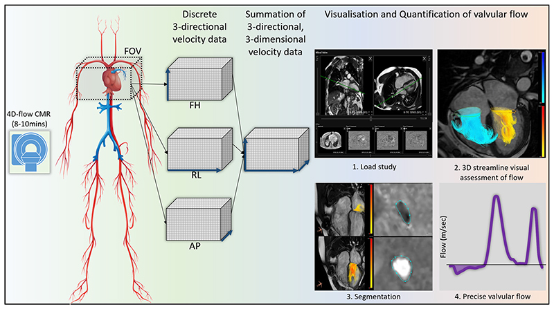
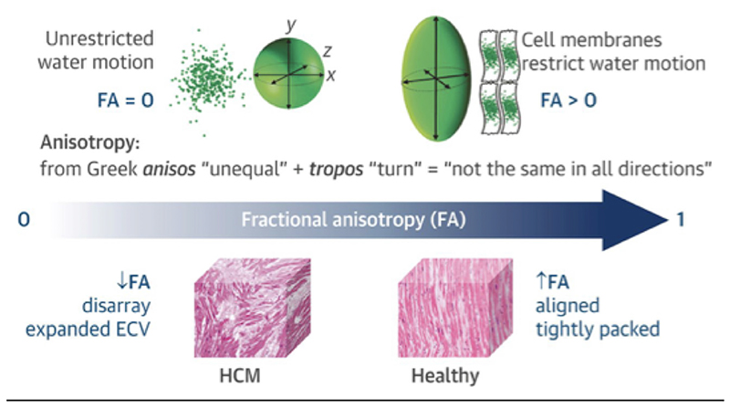
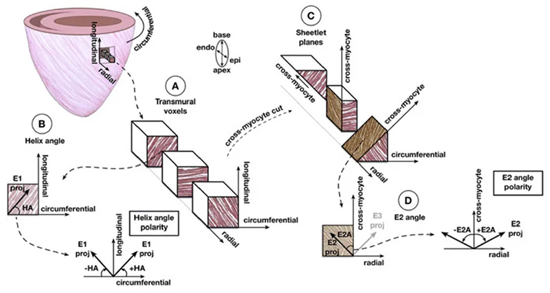
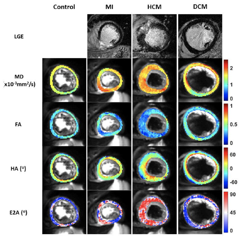
This review gives examples of emerging cardiovascular magnetic resonance (CMR) techniques and applications that have the potential to transition from research to clinical application in the near future. Four-dimensional flow CMR (4D-flow CMR) allows time-resolved three-directional, three-dimensional (3D) velocity-encoded phase-contrast imaging for 3D visualisation and quantification of valvular or intracavity flow. Acquisition times of under 10 min are achievable for a whole heart multidirectional data set and commercial software packages are now available for data analysis, making 4D-flow CMR feasible for inclusion in clinical imaging protocols. Diffusion tensor imaging (DTI) is based on the measurement of molecular water diffusion and uses contrasting behaviour in the presence and absence of boundaries to infer tissue structure. Cardiac DTI is capable of non-invasively phenotyping the 3D micro-architecture within a few minutes, facilitating transition of the method to clinical protocols. Hybrid positron emission tomography-magnetic resonance (PET-MR) provides quantitative PET measures of biological and pathological processes of the heart combined with anatomical, morphological and functional CMR imaging. Cardiac PET-MR offers opportunities in ischaemic, inflammatory and infiltrative heart disease.
这篇综述介绍了一些新兴的心血管磁共振(CMR)技术和应用,它们有可能在不久的将来从研究转化为临床应用。四维血流 CMR(4D-flow CMR)允许对瓣膜或心腔内的三维(3D)速度编码的相位对比成像进行时间分辨的三维(3D)方向流速测量,用于 3D 可视化和量化。整个心脏多方向数据集的采集时间可在 10 分钟以内完成,并且现在有商业软件包可用于数据分析,这使得 4D-flow CMR 可以纳入临床成像方案。扩散张量成像(DTI)基于分子水扩散的测量,利用边界存在和不存在时的对比行为来推断组织结构。心脏 DTI 能够在几分钟内无创地对 3D 微观结构进行表型分析,从而促进该方法向临床方案的转变。正电子发射断层扫描-磁共振(PET-MR)融合提供了心脏的生物学和病理过程的定量 PET 测量,结合了解剖学、形态学和功能 CMR 成像。心脏 PET-MR 在缺血性、炎症性和浸润性心脏病方面提供了机会。