• 文献检索
  • 文档翻译
  • 深度研究
  • 学术资讯
  • Suppr Zotero 插件Zotero 插件
  • 邀请有礼
  • 套餐&价格
  • 历史记录
应用&插件
Suppr Zotero 插件Zotero 插件浏览器插件Mac 客户端Windows 客户端微信小程序
定价
高级版会员购买积分包购买API积分包
服务
文献检索文档翻译深度研究API 文档MCP 服务
关于我们
关于 Suppr公司介绍联系我们用户协议隐私条款
关注我们

Suppr 超能文献

核心技术专利:CN118964589B侵权必究
粤ICP备2023148730 号-1Suppr @ 2026

文献检索

告别复杂PubMed语法,用中文像聊天一样搜索,搜遍4000万医学文献。AI智能推荐,让科研检索更轻松。

立即免费搜索

文件翻译

保留排版,准确专业,支持PDF/Word/PPT等文件格式,支持 12+语言互译。

免费翻译文档

深度研究

AI帮你快速写综述,25分钟生成高质量综述,智能提取关键信息,辅助科研写作。

立即免费体验

CT成像结合三维最大密度投影重建在三级医院门静脉变异评估中的作用

Role of CT Imaging With Three-Dimensional Maximum Intensity Projection Reconstruction in the Evaluation of Portal Vein Variants at a Tertiary Care Hospital.

作者信息

Asad Ullah Muhammad, Ahmed Muhammad Saad, Hamid Kamran, Ali Muhammad, Shazlee Muhammad Kashif, Darira Jaideep

机构信息

Diagnostic Radiology, Dr. Ziauddin Hospital, Karachi, PAK.

Imaging Services, The Indus Hospital, Karachi, PAK.

出版信息

Cureus. 2020 Nov 28;12(11):e11733. doi: 10.7759/cureus.11733.

DOI:10.7759/cureus.11733
PMID:33403165
原文链接:https://pmc.ncbi.nlm.nih.gov/articles/PMC7773306/
Abstract

INTRODUCTION

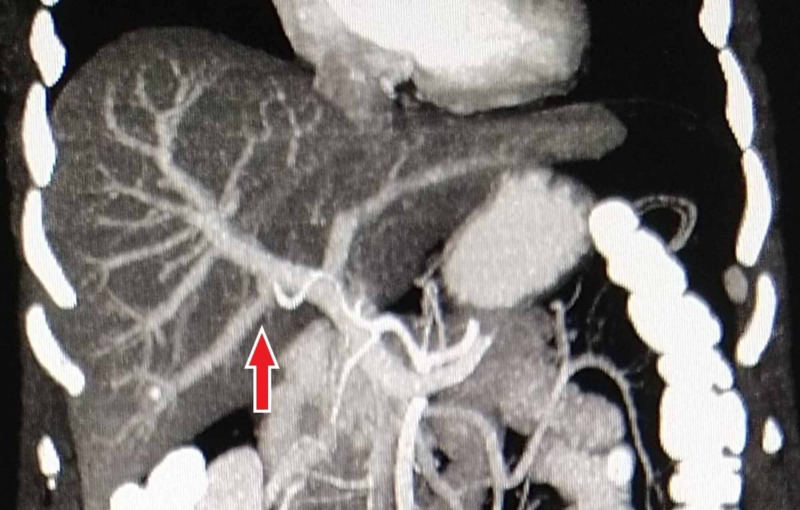
Portal vein (PV) is the principal blood vessel transporting blood from the alimentary tract and spleen to the liver. The aim of this study is to determine the prevalence of PV anatomical variations in our population using multidetector CT with maximum intensity projection (MIP) technique at a tertiary care hospital.

METHODS

This cross-sectional study was prospectively conducted from November 2018 to June 2019 in the Department of Radiology at a tertiary care hospital in Karachi. After informed consent, all the patients with no known hepatic pathology undergoing routine abdomen CT were included in this study. Patients with previous hepatic resection surgeries, undiagnosed large hepatic tumors/metastasis, and those with PV thrombosis were excluded.

RESULTS

A total of 500 patients (256 males and 244 females) were included in the study; the mean age of female patients was relatively higher as compared to the male patients (53.80 ± 18.44 vs. 44.15 ± 19.94 years; p = 0.000). Standard PV anatomy (type 1) was found in 438 patients (87.6%). Trifurcation (type 2) occurred in 18 patients (3.6%). Right posterior portal vein as the first branch of main PV (type 3) was found in 22 patients (4.4%). A separate branch of the right portal vein (RPV) to segment VII (type 4) and separate branch of the RPV to segment VI (type 5) were found in 6 (1.2%) and 16 (3.2%) patients, respectively.

CONCLUSION

Our study displayed a relatively higher frequency of standard PV anatomy (type 1) compared to previous studies. We highlight the role of MIP in the analysis of hepatic venous anatomy with its utility demonstrating improved detection of variations.

摘要

引言

门静脉(PV)是将来自消化道和脾脏的血液输送至肝脏的主要血管。本研究的目的是在一家三级医疗中心医院,使用多排螺旋CT的最大密度投影(MIP)技术确定我们所研究人群中PV解剖变异的发生率。

方法

本横断面研究于2018年11月至2019年6月在卡拉奇一家三级医疗中心医院的放射科前瞻性开展。在获得知情同意后,所有无已知肝脏病变且接受常规腹部CT检查的患者均纳入本研究。既往有肝脏切除手术史、未确诊的大肝脏肿瘤/转移瘤以及PV血栓形成的患者被排除。

结果

本研究共纳入500例患者(男性256例,女性244例);女性患者的平均年龄比男性患者相对更高(53.80±18.44岁 vs. 44.15±19.94岁;p = 0.000)。438例患者(87.6%)发现标准PV解剖(1型)。18例患者(3.6%)出现三叉分支(2型)。22例患者(4.4%)发现右后门静脉作为主PV的第一分支(3型)。分别有6例(1.2%)和16例(3.2%)患者发现右门静脉(RPV)至Ⅶ段的单独分支(4型)和RPV至Ⅵ段的单独分支(5型)。

结论

与既往研究相比,我们的研究显示标准PV解剖(1型)的发生率相对较高。我们强调了MIP在肝静脉解剖分析中的作用,其效用表明能更好地检测变异情况。

https://cdn.ncbi.nlm.nih.gov/pmc/blobs/cdc7/7773306/929448a0ca62/cureus-0012-00000011733-i05.jpg
https://cdn.ncbi.nlm.nih.gov/pmc/blobs/cdc7/7773306/b00ef9a03636/cureus-0012-00000011733-i01.jpg
https://cdn.ncbi.nlm.nih.gov/pmc/blobs/cdc7/7773306/c60ffed7c83e/cureus-0012-00000011733-i02.jpg
https://cdn.ncbi.nlm.nih.gov/pmc/blobs/cdc7/7773306/1e31f7ecadf8/cureus-0012-00000011733-i03.jpg
https://cdn.ncbi.nlm.nih.gov/pmc/blobs/cdc7/7773306/5e879f31af28/cureus-0012-00000011733-i04.jpg
https://cdn.ncbi.nlm.nih.gov/pmc/blobs/cdc7/7773306/929448a0ca62/cureus-0012-00000011733-i05.jpg

相似文献

1
Role of CT Imaging With Three-Dimensional Maximum Intensity Projection Reconstruction in the Evaluation of Portal Vein Variants at a Tertiary Care Hospital.CT成像结合三维最大密度投影重建在三级医院门静脉变异评估中的作用
Cureus. 2020 Nov 28;12(11):e11733. doi: 10.7759/cureus.11733.
2
CT virtual endoscopy for analyzing variations in the hepatic portal vein.用于分析肝门静脉变异的CT虚拟内窥镜检查
Surg Radiol Anat. 2015 Jul;37(5):457-62. doi: 10.1007/s00276-015-1463-2. Epub 2015 Mar 25.
3
Multislice CT angiography in the evaluation of hepatic vascular anatomy in potential right lobe donors.多层螺旋CT血管造影在潜在右叶供体肝脏血管解剖评估中的应用
Diagn Interv Radiol. 2005 Mar;11(1):51-9.
4
Variations of the right branch of hepatic portal vein in children based on three-dimensional simulation technology.基于三维模拟技术的儿童肝门静脉右支变异。
Surg Radiol Anat. 2020 Dec;42(12):1467-1473. doi: 10.1007/s00276-020-02499-3. Epub 2020 May 18.
5
Portal vein variations: clinical implications and frequencies in routine abdominal multidetector CT.门静脉变异:常规腹部多层螺旋CT的临床意义及发生率
Diagn Interv Radiol. 2007 Jun;13(2):75-80.
6
Reappraisal of right portal segmental ramification based on 3-dimensional volume rendering of computed tomography during arterial portography.基于动脉期门静脉造影计算机断层扫描三维容积再现技术对右门静脉分支的重新评估
J Comput Assist Tomogr. 2007 May-Jun;31(3):475-80. doi: 10.1097/01.rct.0000243448.41233.75.
7
Portal vein variations in 1000 patients: surgical and radiological importance.1000例患者的门静脉变异:手术及影像学意义
Br J Radiol. 2015;88(1055):20150326. doi: 10.1259/bjr.20150326. Epub 2015 Aug 18.
8
Surgical procedures for management of right portal venous branching in right lobe living donor liver transplantation.右半肝活体供肝移植中右门静脉分支处理的手术方法
Transplant Proc. 2008 Jun;40(5):1529-33. doi: 10.1016/j.transproceed.2008.02.085.
9
Portal Vein Variations, Clinical Correlation, and Embryological Explanation: A Review Article.门静脉变异、临床关联及胚胎学解释:一篇综述文章
Cureus. 2023 Mar 20;15(3):e36400. doi: 10.7759/cureus.36400. eCollection 2023 Mar.
10
Usefulness of a saline flush for intravenous 3-dimensional computed tomography portography using multidetector-row helical computed tomography.生理盐水冲洗在使用多排螺旋计算机断层扫描进行静脉三维计算机断层扫描门静脉造影中的应用价值。
J Comput Assist Tomogr. 2005 Nov-Dec;29(6):780-5. doi: 10.1097/01.rct.0000184643.27045.87.

引用本文的文献

1
Examining the Branching Patterns of the Hepatis Portae Vena with Computed Tomography Images.利用计算机断层扫描图像研究肝门静脉分支模式。
J Clin Med. 2025 Jul 8;14(14):4835. doi: 10.3390/jcm14144835.
2
A systematic review and meta-analysis: prevalence and clinical implications of anatomical variants of the hepatic portal vein.一项系统评价与荟萃分析:肝门静脉解剖变异的患病率及临床意义
Sci Rep. 2024 Dec 3;14(1):30002. doi: 10.1038/s41598-024-81543-3.
3
Duplicated inferior vena cava-trifurcated portal vein: a rare anatomical variation encountered during Whipple procedure.

本文引用的文献

1
Comparative analysis of three-dimensional volume rendering and maximum intensity projection for preoperative planning in liver cancer.三维容积再现与最大密度投影在肝癌术前规划中的对比分析
Eur J Radiol Open. 2020 Sep 4;7:100259. doi: 10.1016/j.ejro.2020.100259. eCollection 2020.
2
Study of anatomical variations of hepatic vasculature using multidetector computed tomography angiography.多排螺旋 CT 血管成像在肝血管解剖变异研究中的应用。
Surg Radiol Anat. 2020 Dec;42(12):1449-1457. doi: 10.1007/s00276-020-02532-5. Epub 2020 Jul 20.
3
All about portal vein: a pictorial display to anatomy, variants and physiopathology.
重复下腔静脉-门静脉三叉:胰十二指肠切除术过程中遇到的一种罕见解剖变异。
J Surg Case Rep. 2023 Jan 26;2023(1):rjad014. doi: 10.1093/jscr/rjad014. eCollection 2023 Jan.
门静脉全解:门静脉解剖、变异及病理生理学的图文展示
Insights Imaging. 2019 Mar 21;10(1):38. doi: 10.1186/s13244-019-0716-8.
4
Portal vein abnormalities: an imaging review.门静脉异常:影像学综述
Clin Imaging. 2018 Nov-Dec;52:70-78. doi: 10.1016/j.clinimag.2018.07.002. Epub 2018 Jul 6.
5
Unusual Branching Pattern of the Portal Vein: Its Importance.门静脉的异常分支模式:其重要性。
J Clin Diagn Res. 2017 Aug;11(8):TD01-TD02. doi: 10.7860/JCDR/2017/29011.10390. Epub 2017 Aug 1.
6
Anatomic Variations of the Right Portal Vein: Prevalence, Imaging Features, and Implications for Successful Transjugular Intrahepatic Portosystemic Shunt Creation.右门静脉的解剖变异:发生率、影像学特征及其对成功建立经颈静脉肝内门体分流术的影响
J Clin Imaging Sci. 2017 Mar 28;7:14. doi: 10.4103/jcis.JCIS_10_17. eCollection 2017.
7
Portal vein branching order helps in the recognition of anomalous right-sided round ligament: common features and variations in portal vein anatomy.门静脉分支类型有助于识别异常右侧圆韧带:门静脉解剖的常见特征和变异。
Abdom Radiol (NY). 2017 Jul;42(7):1832-1838. doi: 10.1007/s00261-017-1128-1.
8
Surgical Implications of Portal Vein Variations and Liver Segmentations: A Recent Update.门静脉变异和肝段划分的手术意义:最新进展
J Clin Diagn Res. 2017 Feb;11(2):AE01-AE05. doi: 10.7860/JCDR/2017/25028.9453. Epub 2017 Feb 1.
9
Portal vein variations in 1000 patients: surgical and radiological importance.1000例患者的门静脉变异:手术及影像学意义
Br J Radiol. 2015;88(1055):20150326. doi: 10.1259/bjr.20150326. Epub 2015 Aug 18.
10
Expectations from imaging for pre-transplant evaluation of living donor liver transplantation.活体肝移植术前评估影像学检查的期望
World J Radiol. 2014 Sep 28;6(9):693-707. doi: 10.4329/wjr.v6.i9.693.