Ho Joyce L, Konda Anuja, Rahman Jawaria, Harris Elan, Korn Ron, Sabir Aqsa, Bawany Basil, Gulati Rajesh, Harris Gordon J, Boswell William D, Fong Yuman, Rahmanuddin Syed
City of Hope Comprehensive Cancer Center, Duarte, CA, USA.
Riverside Community Hospital, Riverside, CA, USA.
Eur J Radiol Open. 2020 Sep 4;7:100259. doi: 10.1016/j.ejro.2020.100259. eCollection 2020.
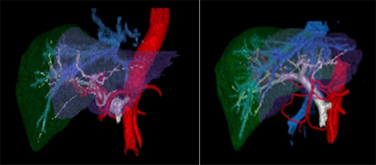
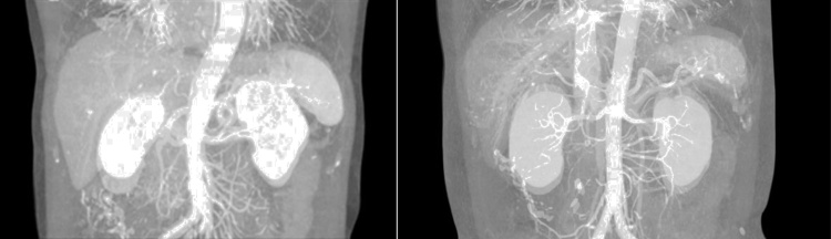
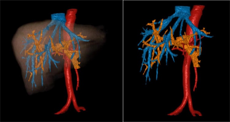
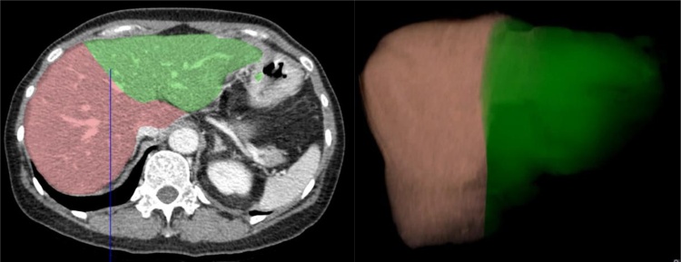
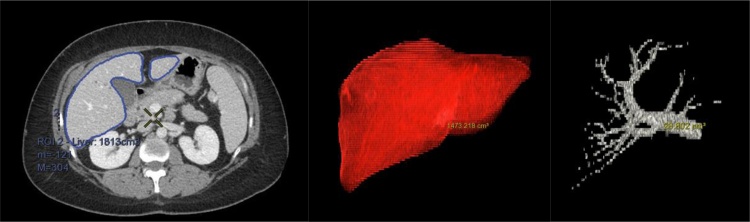
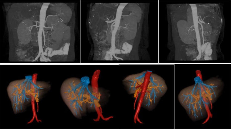
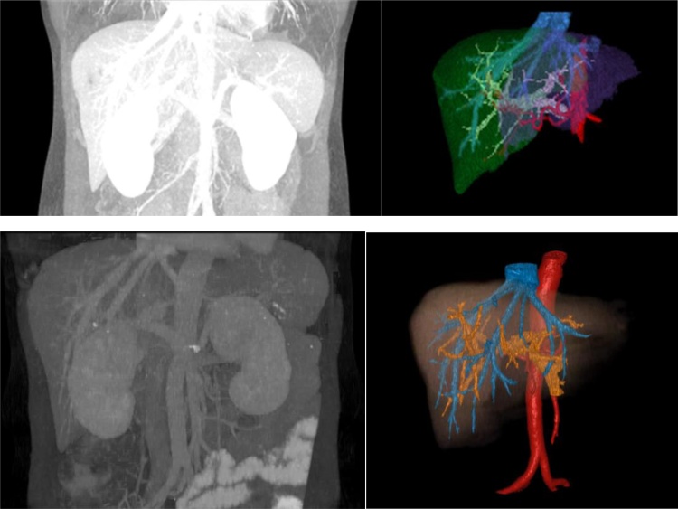
Three-dimensional imaging is a useful tool to evaluate liver structure and surrounding vessels for preoperative planning. In this study, we compared two methods of visualizing vascular maps on computed tomography including maximum intensity projection (MIP) and 3D volume rendered (VR) imaging. We compiled important imaging components of pre-surgical planning, and developed criteria for comparison. The imaging techniques were compared based on colorization, volume quantification, rotation, vessel delineation, small vessel clarity, and segmental liver isolation. MIP had more overall limitations due to reduced differentiation of superimposed structures, motion artifact, and interference from calcifications. We determined that because 3D quantitative volume rendered imaging can provide more detail and perspective than MIP imaging, it may be more useful in preoperative planning for patients with liver malignancy. Advanced 3D imaging is a useful tool that can have profound clinical implications on cancer detection and surgical planning.
三维成像技术是一种用于术前规划中评估肝脏结构及周围血管的有用工具。在本研究中,我们比较了计算机断层扫描上两种血管图谱可视化方法,包括最大密度投影(MIP)和三维容积再现(VR)成像。我们汇总了术前规划的重要成像要素,并制定了比较标准。基于色彩化、容积定量、旋转、血管描绘、小血管清晰度和肝段分离等方面对成像技术进行了比较。由于叠加结构的分辨能力降低、运动伪影以及钙化干扰,MIP存在更多整体局限性。我们确定,由于三维定量容积再现成像比MIP成像能提供更多细节和视角,它在肝恶性肿瘤患者的术前规划中可能更有用。先进的三维成像技术是一种有用工具,对癌症检测和手术规划可能具有深远的临床意义。