Department of Histology and Embryology, Faculty of Medicine, Masaryk University, Brno, Czech Republic.
Reprofit International, Clinic of Reproductive Medicine, Brno, Czech Republic.
Biol Reprod. 2021 Jan 4;104(1):106-116. doi: 10.1093/biolre/ioaa174.
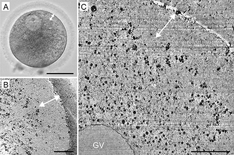
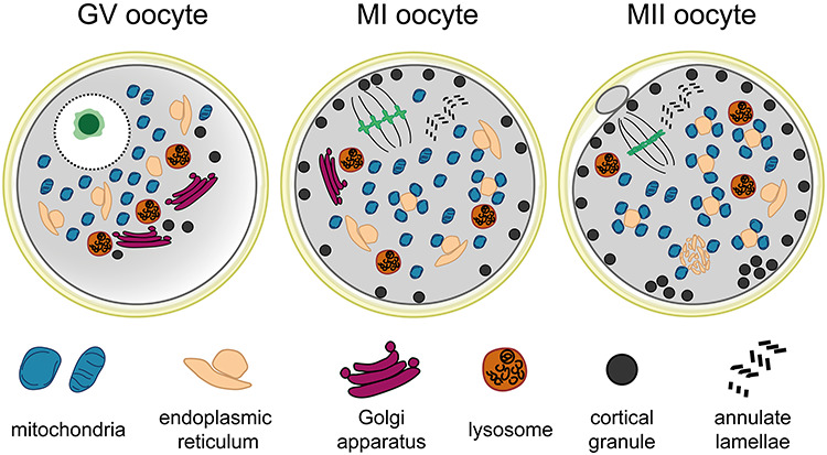
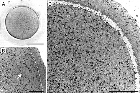
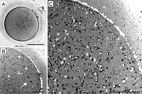
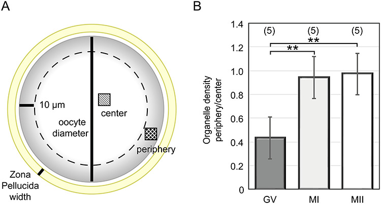
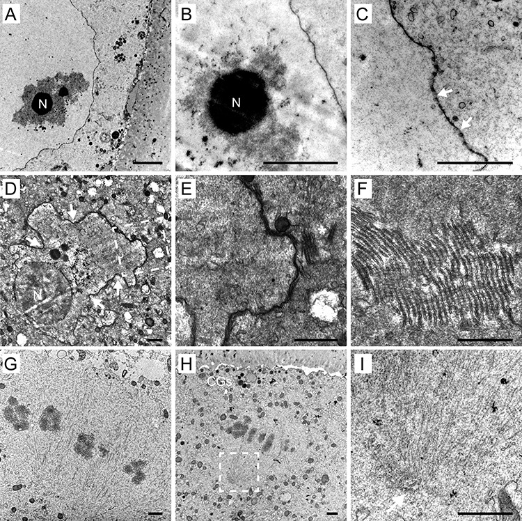
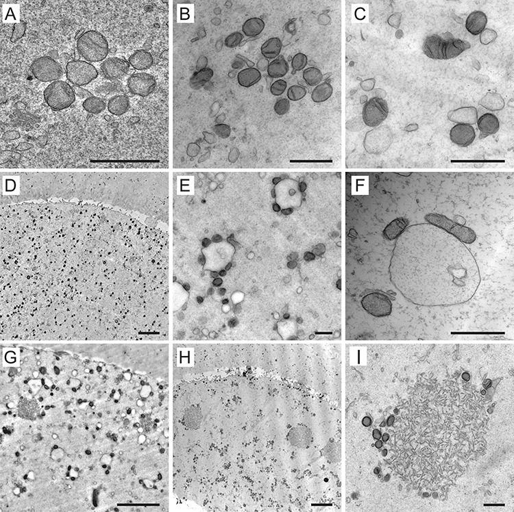
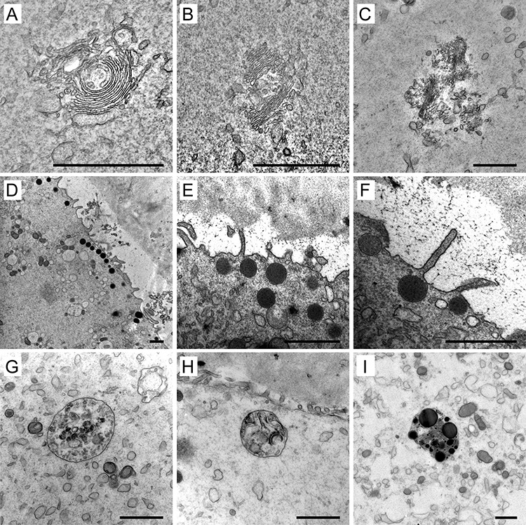
Female fertility relies on successful egg development. Besides chromosome segregation, complex structural and biochemical changes in the cytoplasmic compartment are necessary to confer the female gamete the capacity to undergo normal fertilization and sustain embryonic development. Despite the profound impact on egg quality, morphological bases of cytoplasmic maturation remain largely unknown. Here, we report our findings from the ultrastructural analysis of 69 unfertilized human oocytes from 34 young and healthy egg donors. By comparison of samples fixed at three consecutive developmental stages, we explored how ooplasmic architecture changes during meiotic maturation in vitro. The morphometric image analysis supported observation that the major reorganization of cytoplasm occurs before polar body extrusion. The organelles initially concentrated around prophase nucleus were repositioned toward the periphery and evenly distributed throughout the ooplasm. As maturation progressed, distinct secretory apparatus appeared to transform into cortical granules that clustered underneath the oocyte's surface. The most prominent feature was the gradual formation of heterologous complexes composed of variable elements of endoplasmic reticulum and multiple mitochondria with primitive morphology. Based on the generated image dataset, we proposed a morphological map of cytoplasmic maturation, which may serve as a reference for future comparative studies. In conclusion, this work improves our understanding of human oocyte morphology, cytoplasmic maturation, and intracellular factors defining human egg quality. Although this analysis involved spare oocytes completing development in vitro, it provides essential insight into the enigmatic process by which human egg progenitors prepare for fertilization.
女性的生育能力依赖于卵子的正常发育。除了染色体分离,细胞质隔间的复杂结构和生化变化也是赋予配子正常受精和维持胚胎发育能力的必要条件。尽管细胞质成熟对卵子质量有深远的影响,但细胞质成熟的形态学基础在很大程度上仍然未知。在这里,我们报告了通过对 34 名年轻健康的卵子供体的 69 个未受精人类卵子进行超微结构分析的结果。通过比较在三个连续发育阶段固定的样本,我们探讨了卵母细胞在体外减数分裂成熟过程中细胞质结构如何发生变化。形态计量图像分析支持这样的观察结果,即细胞质的主要重组发生在极体排出之前。最初集中在前期核周围的细胞器被重新定位到细胞质的外围并均匀分布。随着成熟的进行,明显的分泌装置似乎转化为皮质颗粒,这些颗粒聚集在卵子表面的下方。最显著的特征是由内质网和具有原始形态的多个线粒体的可变元件组成的异源复合物的逐渐形成。基于生成的图像数据集,我们提出了细胞质成熟的形态学图谱,该图谱可作为未来比较研究的参考。总之,这项工作提高了我们对人类卵子形态、细胞质成熟以及决定人类卵子质量的细胞内因素的理解。虽然这项分析涉及在体外完成发育的备用卵子,但它为神秘的人类卵子前体为受精做准备的过程提供了重要的见解。