Department of Cardiology, The First Affiliated Hospital of Guangxi Medical University, 6 Shuangyong Road, Nanning, 530021, China.
Department of Cardiology, Fourth Affiliated Hospital of Guangxi Medical University, Liuzhou, China.
BMC Cardiovasc Disord. 2021 Jan 6;21(1):5. doi: 10.1186/s12872-020-01775-9.
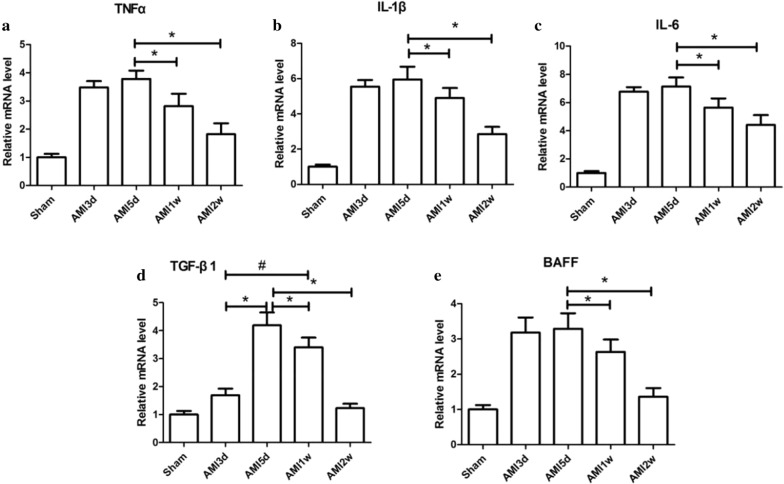
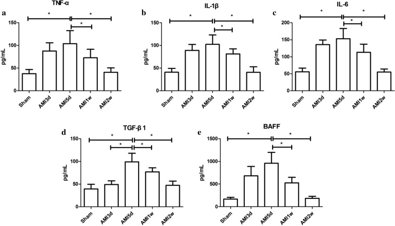
Inflammatory cells infiltrate into the ischemic and hypoxic myocardial tissue after myocardial infarction. B cells gather at the site of myocardial injury and secrete cytokines to regulate immune inflammation and fiber repair processes.
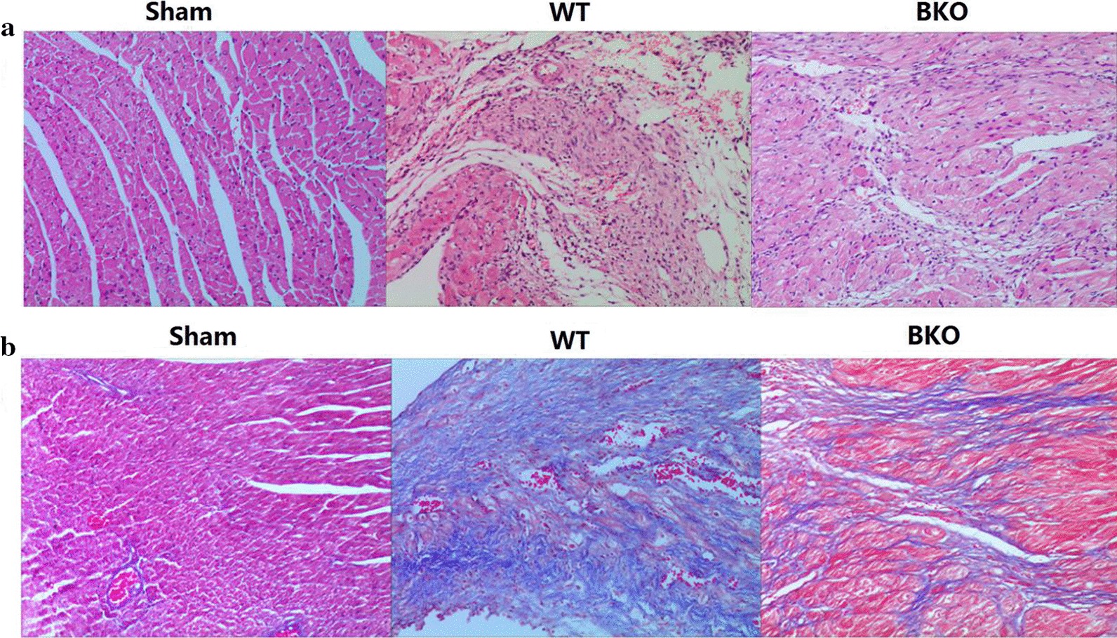
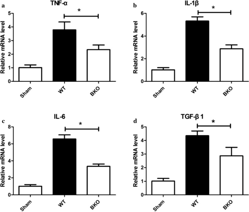
The animal experiment used ligation of the left anterior descending (LAD) artery of C57BL/6 mice to establish a mouse acute myocardial infarction (AMI) model to observe changes in activated B cells and cytokines at different time points. Twelve-week-old C57BL/6 male mice were randomly divided into the Sham group (24 mice) (thread under the LAD artery without ligation) and the AMI group (64 mice). In addition, C57BL/6 B-cell knockout (BKO) mice and C57BL/6 wild-type (WT) mice were used to establish AMI models to observe the expression levels of cardiomyocyte cytokines, such as TNF-α IL-1β, IL-6, TGF-β1, COL1-A1, COL3-AIII, TIMP, and MMP9. Moreover, pathological and collagen changes in the myocardium were analysed. One-way ANOVA and LSD method was used for comparisons of multiple and pairwise groups respectively. P < 0.05 indicated significant differences.
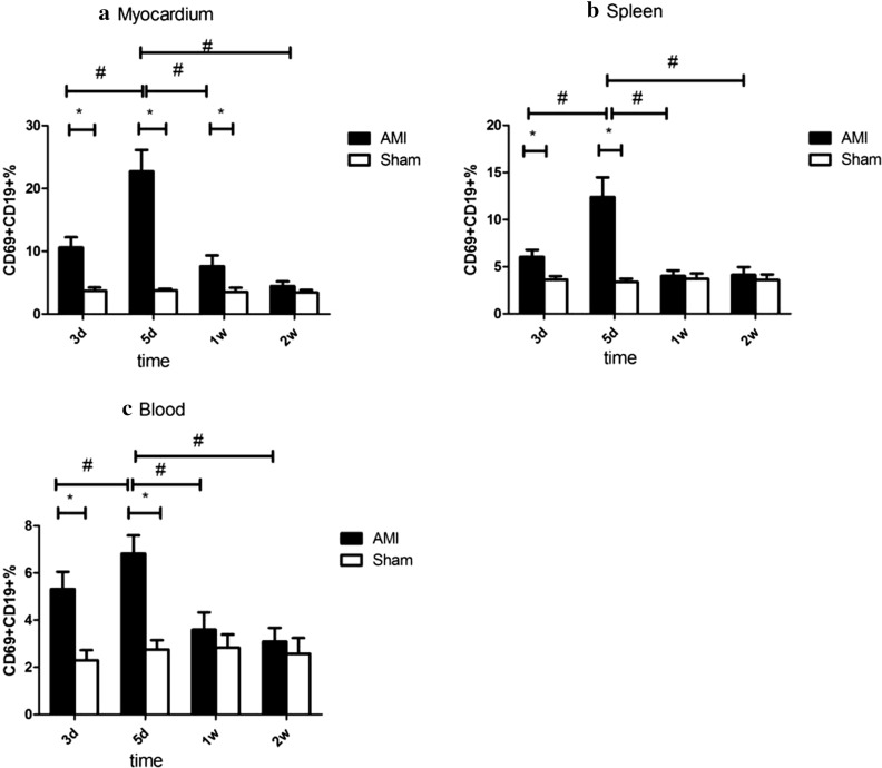
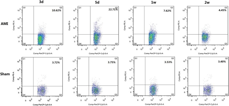
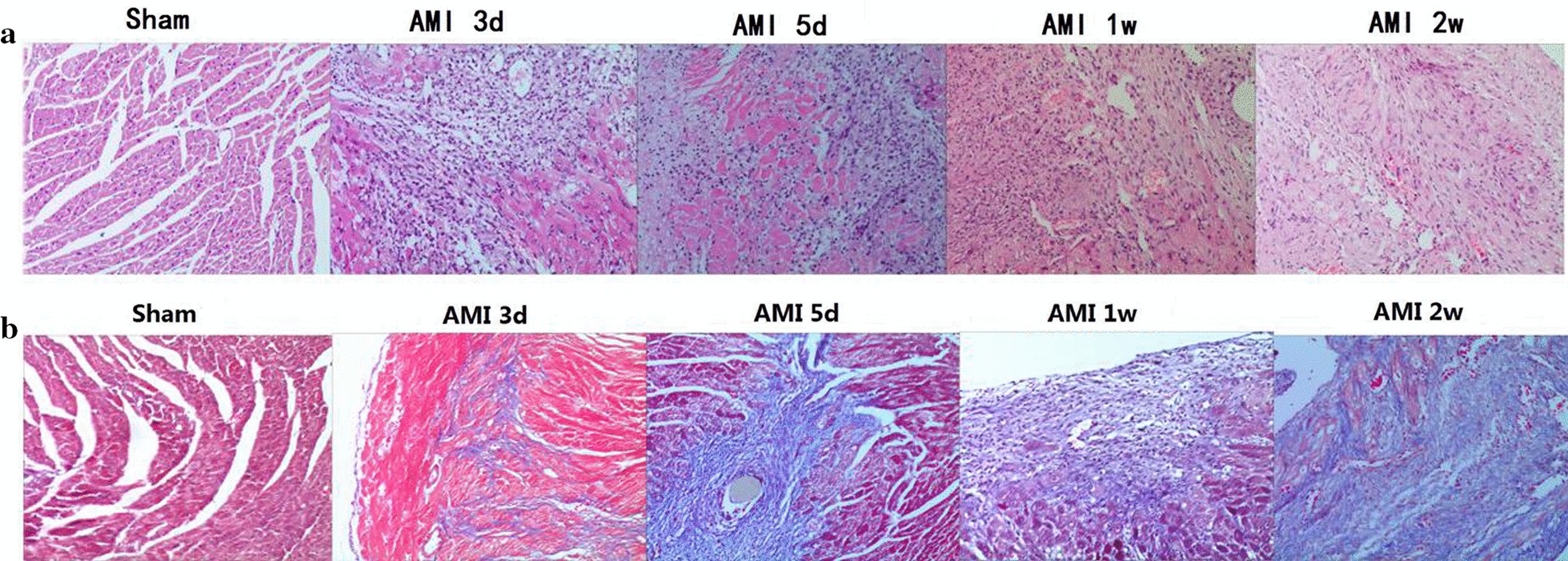
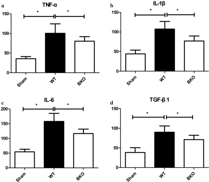
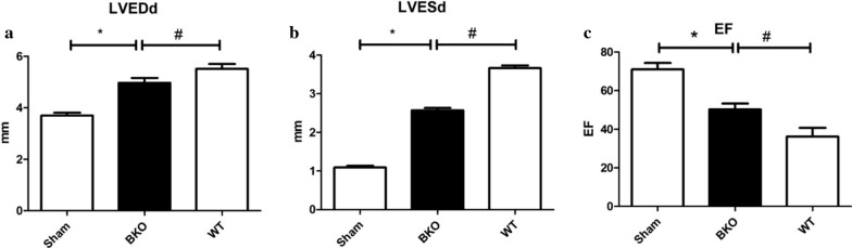
An AMI model of C57BL/6 mice was established successfully. The ratio of activated B cells and the expression of TNF-α, IL-1β, IL-6, TGF-β1, and B cell activating factor (BAFF) in the 5-day subgroup were the highest in the myocardium, spleen and peripheral blood with the most obvious myocardial inflammatory cell infiltration. The cytokines mRNA expression levels in the 5-day subgroup of the BKO group were decreased compared with those in the WT group (P < 0.05). Among the 2-week subgroups of the Sham, WT and BKO groups, the the LVEDd and LVESd of the BKO group were lower than those of the WT group (P < 0.05), and the left ventricular ejection fraction was higher than that of the WT group (P < 0.05).
Activated B cells participate in the sustained state of myocardial inflammation and immune system activation after AMI, and may affect the metabolism of myocardial collagen after AMI by secreting cytokines. Moreover, B cells promote the expression of myocardial collagen Type I and Type III and damage the left ventricular ejection function.
炎症细胞浸润到心肌梗死后的缺血缺氧心肌组织中。B 细胞聚集在心肌损伤部位,分泌细胞因子调节免疫炎症和纤维修复过程。
动物实验采用结扎 C57BL/6 小鼠的左前降支(LAD)动脉建立小鼠急性心肌梗死(AMI)模型,观察不同时间点激活 B 细胞和细胞因子的变化。将 12 周龄的 C57BL/6 雄性小鼠随机分为 Sham 组(24 只)(LAD 动脉下的线未结扎)和 AMI 组(64 只)。此外,还使用 C57BL/6 B 细胞敲除(BKO)小鼠和 C57BL/6 野生型(WT)小鼠建立 AMI 模型,观察 TNF-α、IL-1β、IL-6、TGF-β1、COL1-A1、COL3-AIII、TIMP 和 MMP9 等心肌细胞因子的表达水平。同时分析心肌的病理和胶原变化。采用单因素方差分析和 LSD 法分别进行多组和两组间的比较。P<0.05 表示差异有统计学意义。
成功建立了 C57BL/6 小鼠 AMI 模型。5 天亚组心肌、脾脏和外周血中激活 B 细胞的比例和 TNF-α、IL-1β、IL-6、TGF-β1 和 B 细胞激活因子(BAFF)的表达最高,心肌炎症细胞浸润最明显。与 WT 组相比,BKO 组 5 天亚组的细胞因子 mRNA 表达水平降低(P<0.05)。在 Sham、WT 和 BKO 组的 2 周亚组中,BKO 组的 LVEDd 和 LVESd 低于 WT 组(P<0.05),左心室射血分数高于 WT 组(P<0.05)。
激活的 B 细胞参与 AMI 后心肌炎症和免疫系统的持续激活,并可能通过分泌细胞因子影响 AMI 后心肌胶原的代谢。此外,B 细胞促进心肌胶原 I 型和 III 型的表达,损害左心室射血功能。