• 文献检索
  • 文档翻译
  • 深度研究
  • 学术资讯
  • Suppr Zotero 插件Zotero 插件
  • 邀请有礼
  • 套餐&价格
  • 历史记录
应用&插件
Suppr Zotero 插件Zotero 插件浏览器插件Mac 客户端Windows 客户端微信小程序
定价
高级版会员购买积分包购买API积分包
服务
文献检索文档翻译深度研究API 文档MCP 服务
关于我们
关于 Suppr公司介绍联系我们用户协议隐私条款
关注我们

Suppr 超能文献

核心技术专利:CN118964589B侵权必究
粤ICP备2023148730 号-1Suppr @ 2026

文献检索

告别复杂PubMed语法,用中文像聊天一样搜索,搜遍4000万医学文献。AI智能推荐,让科研检索更轻松。

立即免费搜索

文件翻译

保留排版,准确专业,支持PDF/Word/PPT等文件格式,支持 12+语言互译。

免费翻译文档

深度研究

AI帮你快速写综述,25分钟生成高质量综述,智能提取关键信息,辅助科研写作。

立即免费体验

用于子宫内膜病变分类的深度学习模型。

Deep learning model for classifying endometrial lesions.

作者信息

Zhang YunZheng, Wang ZiHao, Zhang Jin, Wang CuiCui, Wang YuShan, Chen Hao, Shan LuHe, Huo JiaNing, Gu JiaHui, Ma Xiaoxin

机构信息

Department of Obstetrics and Gynecology, Shengjing Hospital of China Medical University, 39 Huaxiang Road, Shenyang, 110021, People's Republic of China.

出版信息

J Transl Med. 2021 Jan 6;19(1):10. doi: 10.1186/s12967-020-02660-x.

DOI:10.1186/s12967-020-02660-x
PMID:33407588
原文链接:https://pmc.ncbi.nlm.nih.gov/articles/PMC7788977/
Abstract

BACKGROUND

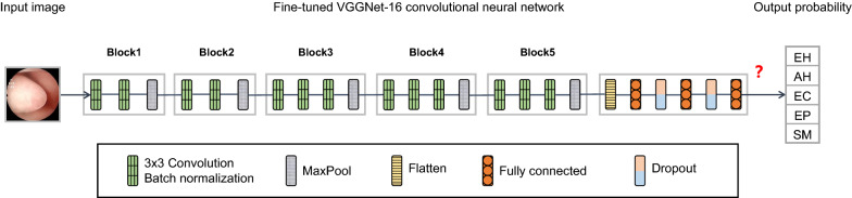
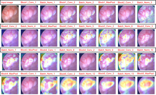
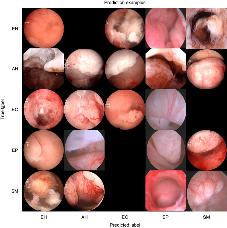
Hysteroscopy is a commonly used technique for diagnosing endometrial lesions. It is essential to develop an objective model to aid clinicians in lesion diagnosis, as each type of lesion has a distinct treatment, and judgments of hysteroscopists are relatively subjective. This study constructs a convolutional neural network model that can automatically classify endometrial lesions using hysteroscopic images as input.

METHODS

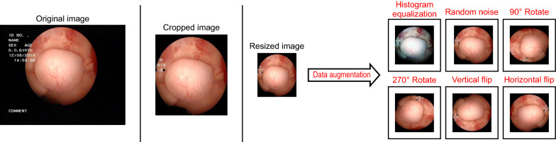
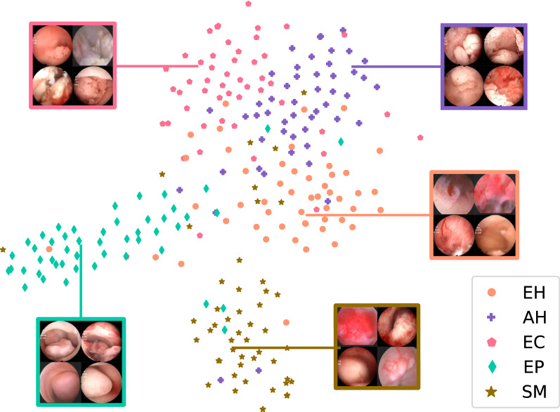
All histopathologically confirmed endometrial lesion images were obtained from the Shengjing Hospital of China Medical University, including endometrial hyperplasia without atypia, atypical hyperplasia, endometrial cancer, endometrial polyps, and submucous myomas. The study included 1851 images from 454 patients. After the images were preprocessed (histogram equalization, addition of noise, rotations, and flips), a training set of 6478 images was input into a tuned VGGNet-16 model; 250 images were used as the test set to evaluate the model's performance. Thereafter, we compared the model's results with the diagnosis of gynecologists.

RESULTS

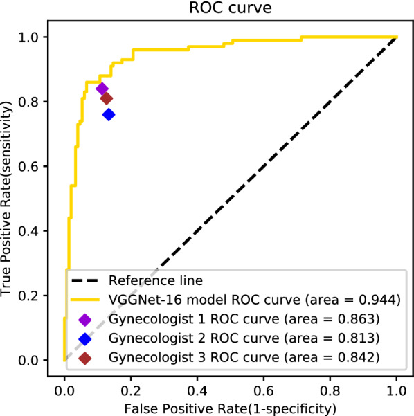
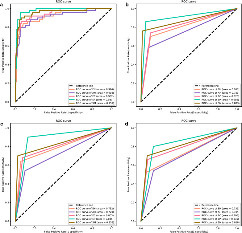
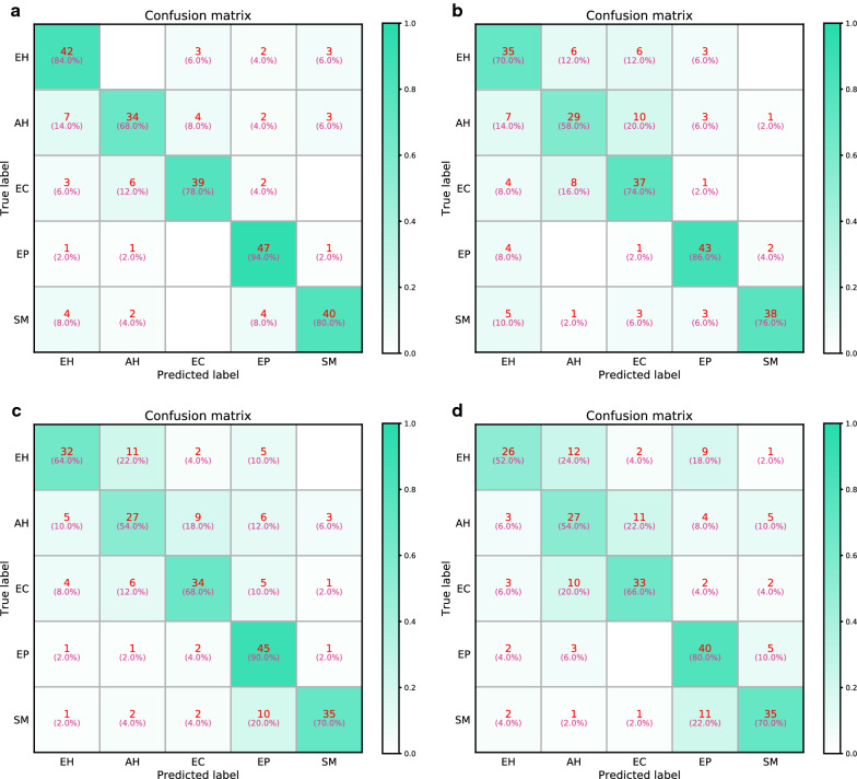
The overall accuracy of the VGGNet-16 model in classifying endometrial lesions is 80.8%. Its sensitivity to endometrial hyperplasia without atypia, atypical hyperplasia, endometrial cancer, endometrial polyp, and submucous myoma is 84.0%, 68.0%, 78.0%, 94.0%, and 80.0%, respectively; for these diagnoses, the model's specificity is 92.5%, 95.5%, 96.5%, 95.0%, and 96.5%, respectively. When classifying lesions as benign or as premalignant/malignant, the VGGNet-16 model's accuracy, sensitivity, and specificity are 90.8%, 83.0%, and 96.0%, respectively. The diagnostic performance of the VGGNet-16 model is slightly better than that of the three gynecologists in both classification tasks. With the aid of the model, the overall accuracy of the diagnosis of endometrial lesions by gynecologists can be improved.

CONCLUSIONS

The VGGNet-16 model performs well in classifying endometrial lesions from hysteroscopic images and can provide objective diagnostic evidence for hysteroscopists.

摘要

背景

宫腔镜检查是诊断子宫内膜病变的常用技术。由于每种病变类型都有独特的治疗方法,且宫腔镜检查医生的判断相对主观,因此开发一种客观模型以协助临床医生进行病变诊断至关重要。本研究构建了一种卷积神经网络模型,该模型可以将宫腔镜图像作为输入自动对子宫内膜病变进行分类。

方法

所有经组织病理学确诊的子宫内膜病变图像均来自中国医科大学盛京医院,包括无异型性的子宫内膜增生、不典型增生、子宫内膜癌、子宫内膜息肉和黏膜下肌瘤。该研究纳入了来自454例患者的1851张图像。在对图像进行预处理(直方图均衡化、添加噪声、旋转和翻转)后,将6478张图像的训练集输入到经过调优的VGGNet-16模型中;250张图像用作测试集以评估模型的性能。此后,我们将模型的结果与妇科医生的诊断进行了比较。

结果

VGGNet-16模型对子宫内膜病变进行分类的总体准确率为80.8%。其对无异型性的子宫内膜增生、不典型增生、子宫内膜癌、子宫内膜息肉和黏膜下肌瘤的敏感性分别为84.0%、68.0%、78.0%、94.0%和80.0%;对于这些诊断,模型的特异性分别为92.5%、95.5%、96.5%、95.0%和96.5%。当将病变分类为良性或癌前/恶性时,VGGNet-16模型的准确率、敏感性和特异性分别为90.8%、83.0%和96.0%。在两项分类任务中,VGGNet-16模型的诊断性能均略优于三位妇科医生。借助该模型,可以提高妇科医生对子宫内膜病变诊断的总体准确率。

结论

VGGNet-16模型在对宫腔镜图像中的子宫内膜病变进行分类方面表现良好,可为宫腔镜检查医生提供客观的诊断依据。

https://cdn.ncbi.nlm.nih.gov/pmc/blobs/40d2/7788977/e22b76460c56/12967_2020_2660_Fig9_HTML.jpg
https://cdn.ncbi.nlm.nih.gov/pmc/blobs/40d2/7788977/10813ce3d0b8/12967_2020_2660_Fig1_HTML.jpg
https://cdn.ncbi.nlm.nih.gov/pmc/blobs/40d2/7788977/4bc9ec52224f/12967_2020_2660_Fig2_HTML.jpg
https://cdn.ncbi.nlm.nih.gov/pmc/blobs/40d2/7788977/9f6af557f030/12967_2020_2660_Fig3_HTML.jpg
https://cdn.ncbi.nlm.nih.gov/pmc/blobs/40d2/7788977/fc712386e6c1/12967_2020_2660_Fig4_HTML.jpg
https://cdn.ncbi.nlm.nih.gov/pmc/blobs/40d2/7788977/310e39f40211/12967_2020_2660_Fig5_HTML.jpg
https://cdn.ncbi.nlm.nih.gov/pmc/blobs/40d2/7788977/0d8390c54da9/12967_2020_2660_Fig6_HTML.jpg
https://cdn.ncbi.nlm.nih.gov/pmc/blobs/40d2/7788977/7f83b52113c0/12967_2020_2660_Fig7_HTML.jpg
https://cdn.ncbi.nlm.nih.gov/pmc/blobs/40d2/7788977/103a94e0b149/12967_2020_2660_Fig8_HTML.jpg
https://cdn.ncbi.nlm.nih.gov/pmc/blobs/40d2/7788977/e22b76460c56/12967_2020_2660_Fig9_HTML.jpg

相似文献

1
Deep learning model for classifying endometrial lesions.用于子宫内膜病变分类的深度学习模型。
J Transl Med. 2021 Jan 6;19(1):10. doi: 10.1186/s12967-020-02660-x.
2
Hysteroscopic Photodynamic Diagnosis Using 5-Aminolevulinic Acid: A High-Sensitivity Diagnostic Method for Uterine Endometrial Malignant Diseases.宫腔镜下 5-氨基酮戊酸光动力诊断:一种用于子宫子宫内膜恶性疾病的高灵敏度诊断方法。
J Minim Invasive Gynecol. 2020 Jul-Aug;27(5):1087-1094. doi: 10.1016/j.jmig.2019.08.012. Epub 2019 Aug 12.
3
Automated system for diagnosing endometrial cancer by adopting deep-learning technology in hysteroscopy.采用深度学习技术的宫腔镜子宫内膜癌自动诊断系统。
PLoS One. 2021 Mar 31;16(3):e0248526. doi: 10.1371/journal.pone.0248526. eCollection 2021.
4
Hysteroscopy in women with abnormal uterine bleeding: a meta-analysis on four major endometrial pathologies.子宫异常出血女性的宫腔镜检查:四种主要子宫内膜病变的荟萃分析
Arch Gynecol Obstet. 2015 Jun;291(6):1347-54. doi: 10.1007/s00404-014-3585-x. Epub 2014 Dec 19.
5
[Validity of hysteroscopy in clinical setting: single centre analysis of 605 consecutive hysteroscopies].[宫腔镜检查在临床环境中的有效性:605例连续宫腔镜检查的单中心分析]
Ceska Gynekol. 2008 Dec;73(6):365-9.
6
[Hysteroscopic evaluation of the uterine cavity in postmenopausal women with uterine bleeding].[绝经后子宫出血妇女宫腔的宫腔镜评估]
Ginekol Pol. 2003 Sep;74(9):892-6.
7
The value of hysteroscopic exploration for abnormal uterine bleeding.宫腔镜检查对异常子宫出血的价值。
J Am Assoc Gynecol Laparosc. 1997 Aug;4(4):453-6. doi: 10.1016/s1074-3804(05)80038-6.
8
Construction of deep learning-based convolutional neural network model for automatic detection of fluid hysteroscopic endometrial micropolyps in infertile women with chronic endometritis.基于深度学习的卷积神经网络模型构建,用于自动检测慢性子宫内膜炎不孕妇女宫腔镜下子宫内膜微息肉。
Eur J Obstet Gynecol Reprod Biol. 2024 Jun;297:249-253. doi: 10.1016/j.ejogrb.2024.04.026. Epub 2024 Apr 21.
9
Detection of benign intracavitary lesions in postmenopausal women with abnormal uterine bleeding: a prospective comparative study on outpatient hysteroscopy and blind biopsy.绝经后异常子宫出血妇女良性腔内病变的检测:门诊宫腔镜检查与盲目活检的前瞻性比较研究
J Minim Invasive Gynecol. 2008 Jan-Feb;15(1):87-91. doi: 10.1016/j.jmig.2007.10.014.
10
Suspecting malignancy in endometrial polyps: value of hysteroscopy.子宫内膜息肉恶性病变的可疑性:宫腔镜检查的价值
Tumori. 2013 Mar-Apr;99(2):204-9. doi: 10.1177/030089161309900214.

引用本文的文献

1
Artificial Intelligence and Hysteroscopy: A Multicentric Study on Automated Classification of Pleomorphic Lesions.人工智能与宫腔镜检查:多中心多形性病变自动分类研究
Cancers (Basel). 2025 Aug 3;17(15):2559. doi: 10.3390/cancers17152559.
2
A deep learning-based computer-aided diagnosis system for detecting atypical endometrial hyperplasia and endometrial cancer through hysteroscopy.一种基于深度学习的计算机辅助诊断系统,用于通过宫腔镜检查检测非典型子宫内膜增生和子宫内膜癌。
iScience. 2025 Jul 3;28(8):113045. doi: 10.1016/j.isci.2025.113045. eCollection 2025 Aug 15.
3
Anthropometric Measurements for Predicting Low Appendicular Lean Mass Index for the Diagnosis of Sarcopenia: A Machine Learning Model.

本文引用的文献

1
Automatic Recognition of Laryngoscopic Images Using a Deep-Learning Technique.使用深度学习技术自动识别喉镜图像。
Laryngoscope. 2020 Nov;130(11):E686-E693. doi: 10.1002/lary.28539. Epub 2020 Feb 18.
2
Support System of Cystoscopic Diagnosis for Bladder Cancer Based on Artificial Intelligence.基于人工智能的膀胱癌膀胱镜诊断支持系统。
J Endourol. 2020 Mar;34(3):352-358. doi: 10.1089/end.2019.0509. Epub 2020 Jan 14.
3
Application of deep learning to the classification of uterine cervical squamous epithelial lesion from colposcopy images.
用于预测低四肢瘦体重指数以诊断肌肉减少症的人体测量学指标:一种机器学习模型
J Funct Morphol Kinesiol. 2025 Jul 17;10(3):276. doi: 10.3390/jfmk10030276.
4
AI-Augmented Advances in the Diagnostic Approaches to Endometrial Cancer.人工智能辅助下子宫内膜癌诊断方法的进展
Cancers (Basel). 2025 May 28;17(11):1810. doi: 10.3390/cancers17111810.
5
The application of ultrasound artificial intelligence in the diagnosis of endometrial diseases: Current practice and future development.超声人工智能在子宫内膜疾病诊断中的应用:当前实践与未来发展
Digit Health. 2025 May 14;11:20552076241310060. doi: 10.1177/20552076241310060. eCollection 2025 Jan-Dec.
6
Enhancing clinical decision-making in endometrial cancer through deep learning technology: A review of current research.通过深度学习技术增强子宫内膜癌的临床决策:当前研究综述
Digit Health. 2024 Nov 18;10:20552076241297053. doi: 10.1177/20552076241297053. eCollection 2024 Jan-Dec.
7
Quantitative Assessment of Polarization and Elastic Properties of Endometrial Tissue for Precancer/Cancer Diagnostics Using Multimodal Optical Coherence Tomography.使用多模态光学相干断层扫描对子宫内膜组织的极化和弹性特性进行定量评估以用于癌前/癌症诊断
Diagnostics (Basel). 2024 Sep 25;14(19):2131. doi: 10.3390/diagnostics14192131.
8
Hysteroscopy: where did we start, and where are we now? The compelling story of what many considered the "Cinderella" of gynecological endoscopy.宫腔镜检查:我们从哪里开始,现在又在哪里?这是一个引人入胜的故事,讲述了许多人认为的妇科内窥镜检查中的“灰姑娘”。
Arch Gynecol Obstet. 2024 Oct;310(4):1877-1888. doi: 10.1007/s00404-024-07677-x. Epub 2024 Aug 16.
9
Bridging the Diagnostic Gap between Histopathologic and Hysteroscopic Chronic Endometritis with Deep Learning Models.利用深度学习模型弥合组织病理学和宫腔镜慢性子宫内膜炎之间的诊断差距。
Medicina (Kaunas). 2024 Jun 12;60(6):972. doi: 10.3390/medicina60060972.
10
Development of an Automated Morphometric Approach to Assess Vascular Outcomes following Exposure to Environmental Chemicals in Zebrafish.开发一种自动形态测量方法以评估斑马鱼暴露于环境化学物质后的血管结局。
Environ Health Perspect. 2024 May;132(5):57001. doi: 10.1289/EHP13214. Epub 2024 May 3.
深度学习在阴道镜图像子宫颈鳞状上皮病变分类中的应用。
Mol Clin Oncol. 2019 Dec;11(6):583-589. doi: 10.3892/mco.2019.1932. Epub 2019 Oct 4.
4
Automated classification of gastric neoplasms in endoscopic images using a convolutional neural network.使用卷积神经网络对内镜图像中的胃肿瘤进行自动分类。
Endoscopy. 2019 Dec;51(12):1121-1129. doi: 10.1055/a-0981-6133. Epub 2019 Aug 23.
5
Identification of cecum time-location in a colonoscopy video by deep learning analysis of colonoscope movement.通过结肠镜运动的深度学习分析识别结肠镜检查视频中盲肠的时间位置
PeerJ. 2019 Jul 29;7:e7256. doi: 10.7717/peerj.7256. eCollection 2019.
6
Modeling Depression Symptoms from Social Network Data through Multiple Instance Learning.通过多实例学习从社交网络数据中建模抑郁症状
AMIA Jt Summits Transl Sci Proc. 2019 May 6;2019:44-53. eCollection 2019.
7
Segmentation of the uterine wall by an ensemble of fully convolutional neural networks.基于全卷积神经网络集成的子宫壁分割
Annu Int Conf IEEE Eng Med Biol Soc. 2018 Jul;2018:49-52. doi: 10.1109/EMBC.2018.8512245.
8
Multimodal Machine Learning: A Survey and Taxonomy.多模态机器学习:一项综述与分类法
IEEE Trans Pattern Anal Mach Intell. 2019 Feb;41(2):423-443. doi: 10.1109/TPAMI.2018.2798607. Epub 2018 Jan 25.
9
Digital Image Analysis with Fully Connected Convolutional Neural Network to Facilitate Hysteroscopic Fibroid Resection.
Gynecol Obstet Invest. 2018;83(6):615-619. doi: 10.1159/000490563. Epub 2018 Jul 5.
10
Comparison of endometrial assessment by transvaginal ultrasonography and hysteroscopy.经阴道超声与宫腔镜检查评估子宫内膜的比较。
Int J Gynaecol Obstet. 2018 Oct;143(1):32-36. doi: 10.1002/ijgo.12567. Epub 2018 Jul 4.