Neural Circuit Development and Regeneration Research Group, Department of Biology, University of Leuven (KU Leuven), Naamsestraat 61, Box 2464, 3000, Leuven, Belgium.
Leuven Brain Institute, Leuven, Belgium.
Acta Neuropathol Commun. 2021 Jan 6;9(1):6. doi: 10.1186/s40478-020-01102-5.
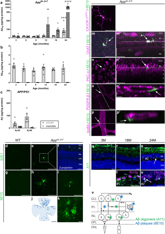
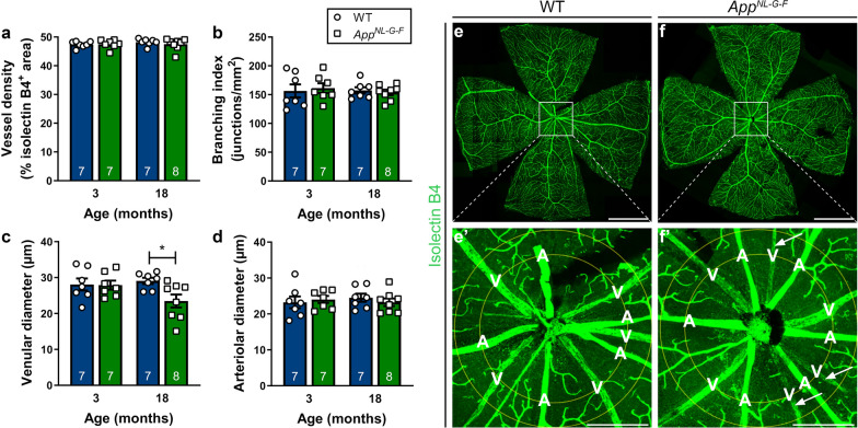
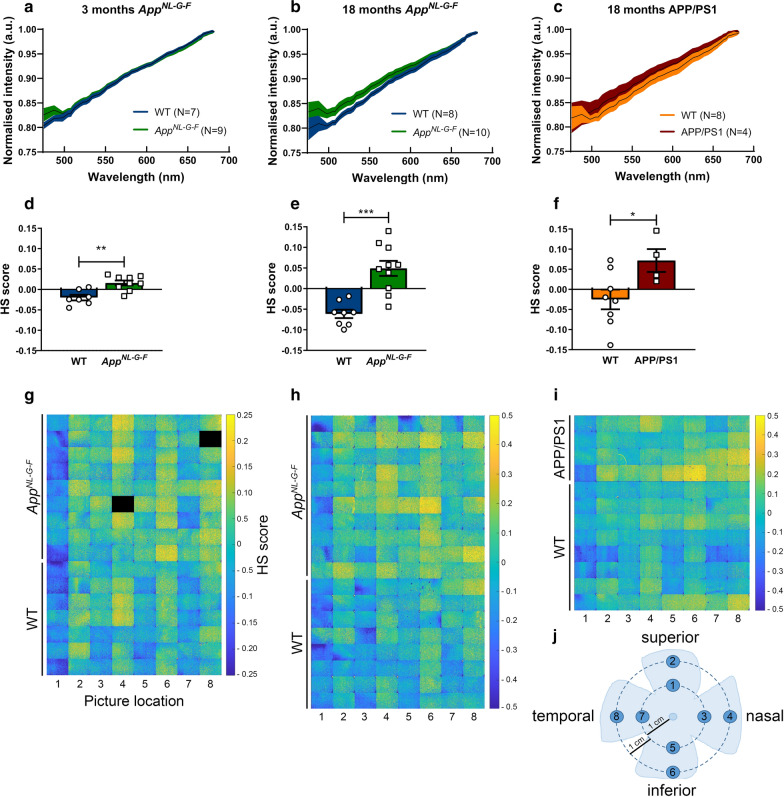
In this study, we report the results of a comprehensive phenotyping of the retina of the App mouse. We demonstrate that soluble Aβ accumulation is present in the retina of these mice early in life and progresses to Aβ plaque formation by midlife. This rising Aβ burden coincides with local microglia reactivity, astrogliosis, and abnormalities in retinal vein morphology. Electrophysiological recordings revealed signs of neuronal dysfunction yet no overt neurodegeneration was observed and visual performance outcomes were unaffected in the App mouse. Furthermore, we show that hyperspectral imaging can be used to quantify retinal Aβ, underscoring its potential as a biomarker for AD diagnosis and monitoring. These findings suggest that the App retina mimics the early, preclinical stages of AD, and, together with retinal imaging techniques, offers unique opportunities for drug discovery and fundamental research into preclinical AD.
在这项研究中,我们报告了 App 小鼠视网膜的全面表型分析结果。我们证明,可溶性 Aβ 在这些小鼠的生命早期就已经在视网膜中积累,并在中年时进展为 Aβ 斑块形成。这种不断增加的 Aβ 负担与局部小胶质细胞反应性、星形胶质细胞增生和视网膜静脉形态异常相吻合。电生理记录显示出神经元功能障碍的迹象,但在 App 小鼠中未观察到明显的神经退行性变,且视觉表现结果不受影响。此外,我们还表明,高光谱成像可用于定量视网膜 Aβ,这突显了它作为 AD 诊断和监测的生物标志物的潜力。这些发现表明,App 视网膜模拟了 AD 的早期、临床前阶段,并且与视网膜成像技术一起,为药物发现和临床前 AD 的基础研究提供了独特的机会。