Harper Danielle J, Augustin Marco, Lichtenegger Antonia, Gesperger Johanna, Himmel Tanja, Muck Martina, Merkle Conrad W, Eugui Pablo, Kummer Stefan, Woehrer Adelheid, Glösmann Martin, Baumann Bernhard
Medical University of Vienna, Center for Medical Physics and Biomedical Engineering, Vienna, Austria.
General Hospital and Medical University of Vienna, Institute of Neurology, Vienna, Austria.
Neurophotonics. 2020 Jan;7(1):015006. doi: 10.1117/1.NPh.7.1.015006. Epub 2020 Feb 4.
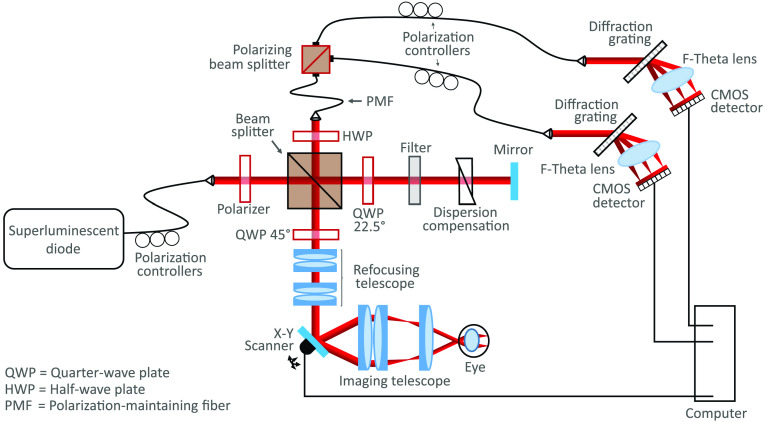
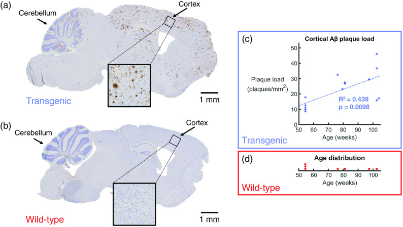
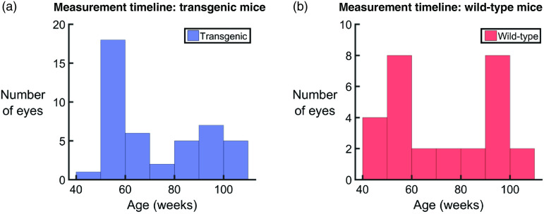
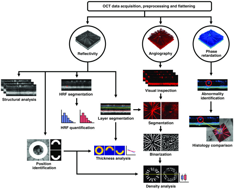
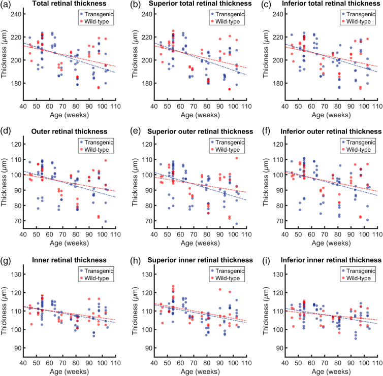
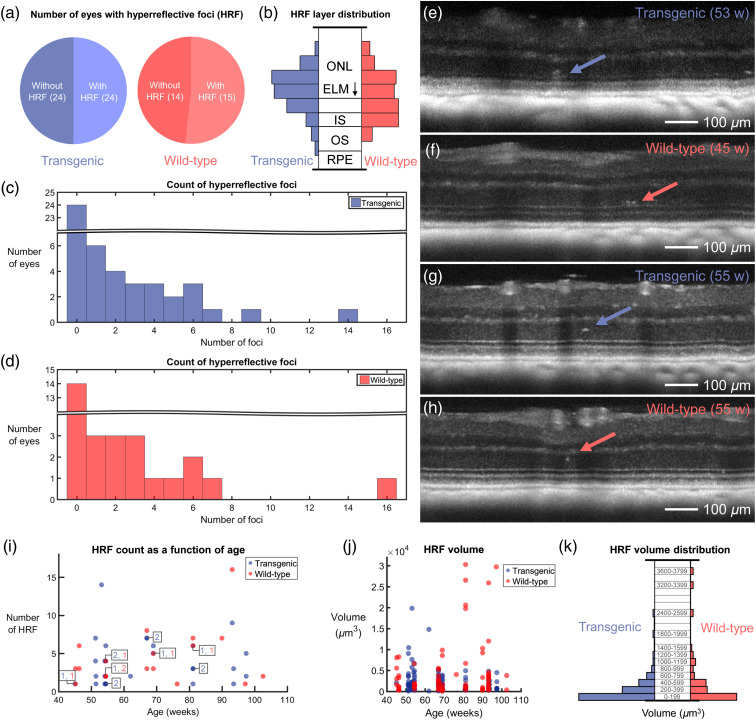
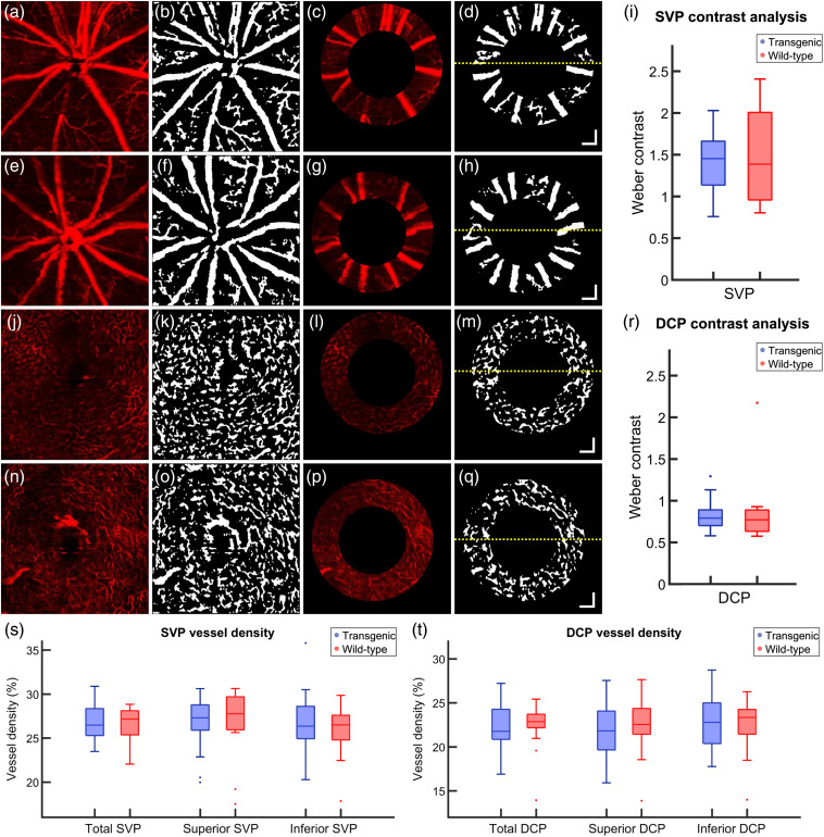
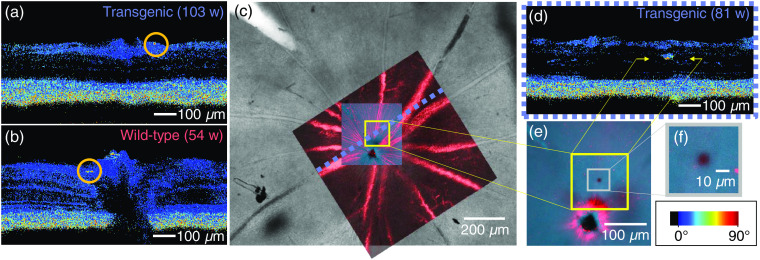
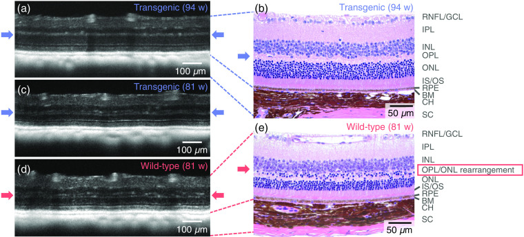
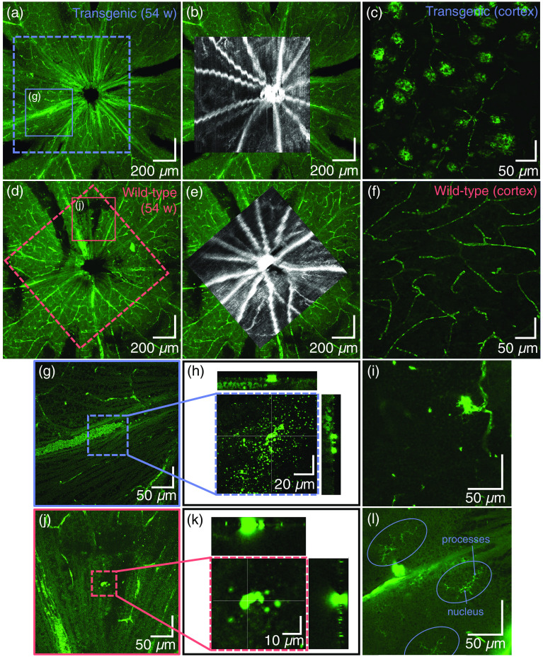
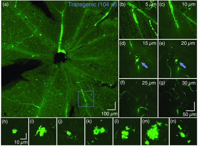
Recent Alzheimer's disease (AD) patient studies have focused on retinal analysis, as the retina is the only part of the central nervous system that can be imaged noninvasively by optical methods. However, as this is a relatively new approach, the occurrence and role of retinal pathological features are still debated. The retina of an APP/PS1 mouse model was investigated using multicontrast optical coherence tomography (OCT) in order to provide a documentation of what was observed in both transgenic and wild-type mice. Both eyes of 24 APP/PS1 transgenic mice (age: 45 to 104 weeks) and 15 age-matched wild-type littermates were imaged by the custom-built OCT system. At the end of the experiment, retinas and brains were harvested from a subset of the mice (14 transgenic, 7 age-matched control) in order to compare the results to histological analysis and to quantify the cortical amyloid beta plaque load. The system provided a combination of standard reflectivity data, polarization-sensitive data, and OCT angiograms. Qualitative and quantitative information from the resultant OCT images was extracted on retinal layer thickness and structure, presence of hyper-reflective foci, phase retardation abnormalities, and retinal vasculature. Although multicontrast OCT revealed abnormal structural properties and phase retardation signals in the retina of this APP/PS1 mouse model, the observations were very similar in transgenic and control mice.
最近针对阿尔茨海默病(AD)患者的研究聚焦于视网膜分析,因为视网膜是中枢神经系统中唯一能够通过光学方法进行无创成像的部分。然而,由于这是一种相对较新的方法,视网膜病理特征的出现情况及作用仍存在争议。为了记录在转基因小鼠和野生型小鼠中观察到的情况,研究人员使用多对比度光学相干断层扫描(OCT)对APP/PS1小鼠模型的视网膜进行了研究。通过定制的OCT系统对24只APP/PS1转基因小鼠(年龄:45至104周)和15只年龄匹配的野生型同窝小鼠的双眼进行了成像。在实验结束时,从一部分小鼠(14只转基因小鼠、7只年龄匹配的对照小鼠)身上采集了视网膜和大脑,以便将结果与组织学分析进行比较,并量化皮质淀粉样β斑块负荷。该系统提供了标准反射率数据、偏振敏感数据和OCT血管造影的组合。从所得的OCT图像中提取了关于视网膜层厚度和结构、高反射灶的存在、相位延迟异常以及视网膜血管系统的定性和定量信息。尽管多对比度OCT显示该APP/PS1小鼠模型的视网膜存在异常结构特性和相位延迟信号,但在转基因小鼠和对照小鼠中的观察结果非常相似。