Eye Institute and Affiliated Xiamen Eye Center of Xiamen University, Fujian, Xiamen, China.
Fujian Provincial Key Laboratory of Ophthalmology and Visual Science, Xiamen, 361102, Fujian, China.
Eye (Lond). 2021 Nov;35(11):3020-3027. doi: 10.1038/s41433-020-01365-1. Epub 2021 Jan 7.
To evaluate the safety and efficacy of repeated corneal collagen crosslinking assisted by transepithelial double-cycle iontophoresis (DI-CXL) in the management of keratoconus progression after primary CXL.
A retrospective analysis was conducted in the patients who underwent repeated CXL between 2016 and 2018. These patients were treated with DI-CXL if keratoconus progression was confirmed after primary CXL. Scoring of ocular pain and corneal epithelial damage, visual acuity, corneal tomography, in vivo corneal confocal microscopy (IVCM) was performed before and at 3, 6, 12, and 24 months after DI-CXL.
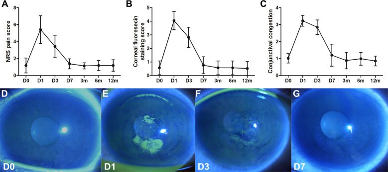
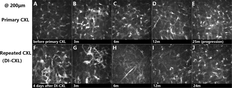
Overall, 21 eyes of 12 patients (mean age 17.3 ± 1.9 years) were included in this study. Before DI-CXL, an average increase of 4.26 D in K was detected in these patients with a mean follow-up interval of (23.0 ± 13.7) months. After DI-CXL, corneal epithelial damage rapidly recovered within days. Visual acuity remained unchanged with follow-up of 24 months. When compared to baseline, significant decreases were observed in K (at 3 months) and K2 (at 3 and 6 months) after DI-CXL. Corneal thickness of thinnest point significantly decreased at 3 months postoperatively. When compared to baseline, no significant differences were found in any of the refractive or tomographic parameters at 12 and 24 months. IVCM revealed trabecular patterned hyperdense tissues after DI-CXL in the anterior stroma at the depth of 200 μm or more. No corneal infiltration or persistent epithelial defect was recorded after DI-CXL.
DI-CXL is safe and effective as a good alternative in stabilizing keratoconus progression after primary CXL.
评估经上皮双循环离子导入(DI-CXL)辅助重复角膜胶原交联治疗初次 CXL 后圆锥角膜进展的安全性和有效性。
对 2016 年至 2018 年期间接受重复 CXL 的患者进行回顾性分析。如果在初次 CXL 后确认圆锥角膜进展,则对这些患者进行 DI-CXL 治疗。在 DI-CXL 前后 3、6、12 和 24 个月进行眼部疼痛和角膜上皮损伤评分、视力、角膜断层扫描、活体角膜共聚焦显微镜(IVCM)检查。
本研究共纳入 12 例患者(平均年龄 17.3±1.9 岁)的 21 只眼。在 DI-CXL 之前,这些患者的 K 值平均增加了 4.26D,平均随访间隔为(23.0±13.7)个月。在 DI-CXL 后,角膜上皮损伤在数天内迅速恢复。视力在 24 个月的随访中保持不变。与基线相比,DI-CXL 后 K(术后 3 个月)和 K2(术后 3 和 6 个月)显著降低。术后 3 个月时角膜最薄点的角膜厚度显著减少。与基线相比,在 12 个月和 24 个月时,任何屈光或断层扫描参数均无显著差异。IVCM 显示 DI-CXL 后在前基质中深度为 200μm 或更深的部位出现小梁状高密度组织。DI-CXL 后未记录到角膜浸润或持续性上皮缺损。
DI-CXL 是一种安全有效的替代方法,可稳定初次 CXL 后圆锥角膜的进展。