Lee Kyuin, Choi Yoon Jung, Choi Hyun Seung, Jeong Junhui
Department of Otorhinolaryngology, National Health Insurance Service Ilsan Hospital, Goyang, Korea.
Department of Pathology, Yongin Severance Hospital, Yonsei University, College of Medicine, Yongin, Korea.
SAGE Open Med Case Rep. 2020 Dec 18;8:2050313X20981469. doi: 10.1177/2050313X20981469. eCollection 2020.
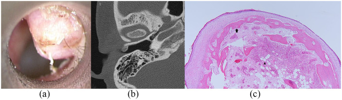
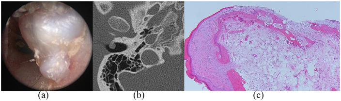
Osteoma of the external auditory canal is a rare benign tumor with an estimated incidence of 0.05% of total otologic surgeries. In most cases, an osteoma in the external auditory canal does not cause symptoms because the tumor grows slowly and does not occlude the ear canal. However, if the mass grows to occlude the external auditory canal, several symptoms can occur, including conductive hearing loss, aural fullness, and keratin debris accumulation. We present two cases of this rare tumor in a 23-year-old woman and a 19-year-old man. The mass was surgically excised at the level of the peduncle under local anesthesia with microscope assistance. The base of the excised mass was drilled with a diamond burr to remove all osseous lesions. Histopathologic findings showed spongiotic osteomas. In these cases, patients had symptoms of aural fullness, although the osteomas did not completely occlude the external auditory canal, and the symptoms improved after surgical excision without recurrence.
外耳道骨瘤是一种罕见的良性肿瘤,在所有耳科手术中的发生率估计为0.05%。在大多数情况下,外耳道骨瘤不会引起症状,因为肿瘤生长缓慢且不会阻塞耳道。然而,如果肿物生长到阻塞外耳道,可能会出现几种症状,包括传导性听力损失、耳闷和角蛋白碎屑积聚。我们报告了两例这种罕见肿瘤的病例,分别为一名23岁女性和一名19岁男性。在显微镜辅助下,于局部麻醉下在蒂部水平手术切除肿物。用金刚石磨头钻除切除肿物的基底部以清除所有骨病变。组织病理学检查结果显示为海绵状骨瘤。在这些病例中,患者有耳闷症状,尽管骨瘤并未完全阻塞外耳道,手术切除后症状改善且无复发。