Department of Biomedical Sciences, Joan C Edwards School of Medicine, Marshall University, Huntington, WV 25755, USA.
Department of Pharmacology and Toxicology, Boonshoft School of Medicine, Wright State University, Dayton, OH 45435, USA.
Oxid Med Cell Longev. 2020 Dec 26;2020:8830537. doi: 10.1155/2020/8830537. eCollection 2020.
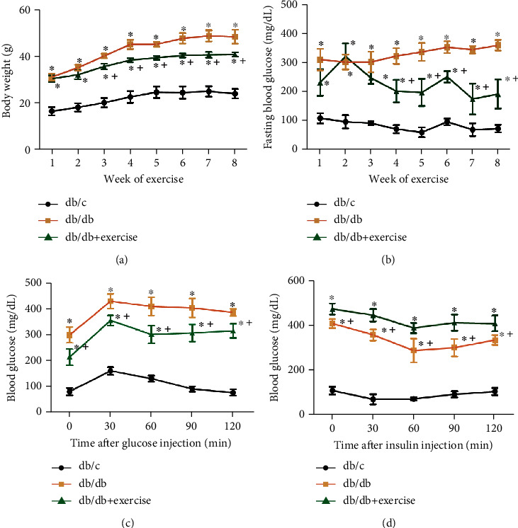
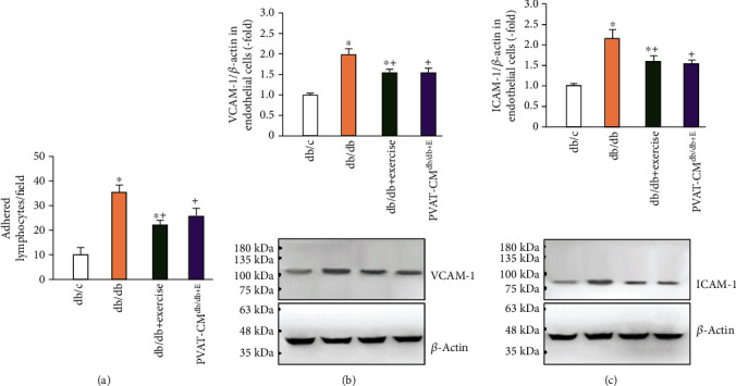
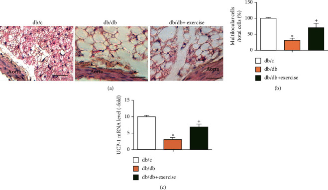
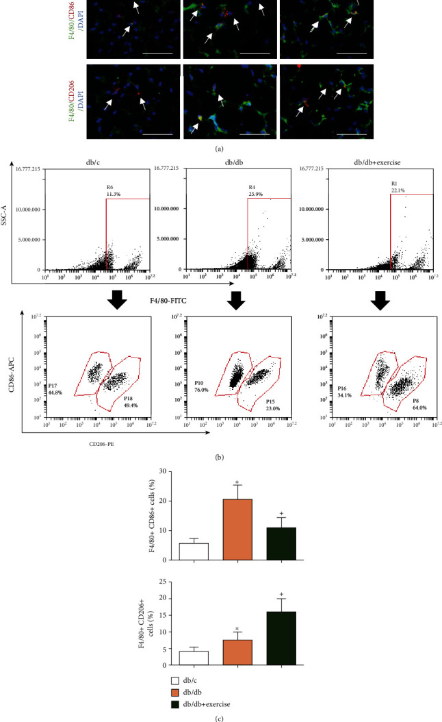
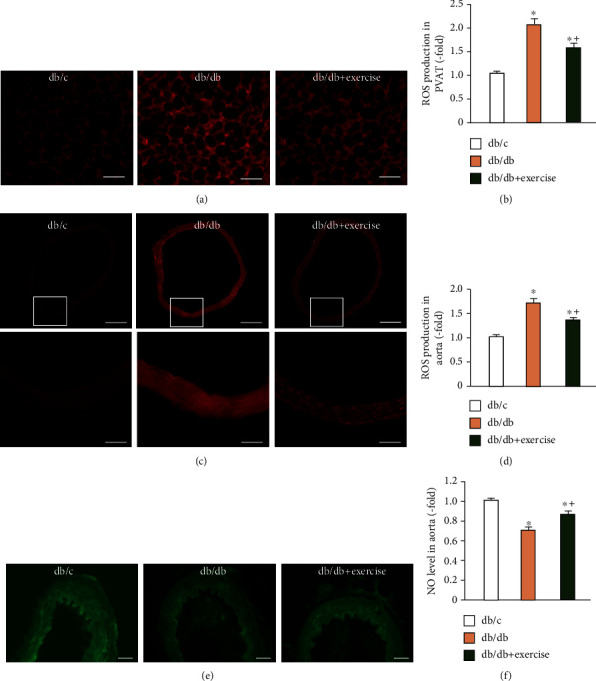
Perivascular adipose tissue (PVAT), a type of adipose tissue that surrounds the blood vessels, has been considered an active component of the blood vessel walls and involved in vascular homeostasis. Recent evidence shows that increased inflammation and oxidative stress in PVAT contribute to endothelial dysfunction in type 2 diabetes (T2D). Exercise is an important nonpharmacological approach for vascular diseases. However, there is limited information regarding whether the beneficial effects of exercise on vascular function is related to the PVAT status. In this study, we investigated whether exercise can decrease oxidative stress and inflammation of PVAT and promote the improvement of endothelial function in a T2D mouse model. Diabetic db/db (5-week old) mice performed treadmill exercise (10 m/min) or keep sedentary for 8 weeks. Body weight, fasting blood glucose levels, glucose, and insulin tolerance were determined. The cytokines (IL-6, IL-10, IFN-, and TNF-a) and adiponectin levels, macrophage polarization and adipocyte type in PVAT, oxidative stress, and nitric oxide (NO) expression in the vascular wall were evaluated. The adhesion ability of primary aorta endothelial cells was analyzed. Our data showed that (1) diabetic db/db mice had increased body weight and fasting blood glucose level, compromised glucose tolerance, and insulin sensitivity, which were decreased/improved by exercise intervention. (2) Exercise intervention increased the percentage of multilocular brown adipocytes, promoted M1 to M2 macrophage polarization, associating with an increase of adiponectin and IL-10 levels and decrease of IFN-, IL-6, and TNF-a levels in PVAT. (3) Exercise decreased superoxide production in PVAT and the vascular wall of diabetic mice, accompanied with increased NO level. (4) The adhesion ability of aorta endothelial cells to leukocytes was decreased in exercised db/db mice, accompanied by decreased intercellular adhesion molecule 1 (ICAM-1) and vascular cell adhesion molecule 1 (VCAM-1) expressions. Of interesting, coculture with PVAT-culture medium from exercised db/db mice could also reduce ICAM-1 and VCAM-1 expressions in primary endothelial cells. In conclusion, our data suggest that exercise improved endothelial function by attenuating the inflammation and oxidative stress in PVAT.
血管周脂肪组织(PVAT)是一种围绕血管的脂肪组织,被认为是血管壁的一个活跃组成部分,参与血管内稳态的调节。最近的证据表明,PVAT 中的炎症和氧化应激增加导致 2 型糖尿病(T2D)中的内皮功能障碍。运动是血管疾病的一种重要非药物治疗方法。然而,关于运动对血管功能的有益作用是否与 PVAT 状态有关,信息有限。在这项研究中,我们研究了运动是否可以降低 T2D 小鼠模型中 PVAT 的氧化应激和炎症,并促进内皮功能的改善。5 周龄的糖尿病 db/db 小鼠进行跑步机运动(10 m/min)或保持久坐 8 周。测定体重、空腹血糖水平、葡萄糖和胰岛素耐量。评估 PVAT 中的细胞因子(IL-6、IL-10、IFN-γ 和 TNF-α)和脂联素水平、巨噬细胞极化和脂肪细胞类型、氧化应激以及血管壁中一氧化氮(NO)的表达。分析原代主动脉内皮细胞的黏附能力。我们的数据表明:(1)糖尿病 db/db 小鼠体重增加和空腹血糖水平升高,葡萄糖耐量和胰岛素敏感性降低,运动干预可降低/改善。(2)运动干预增加了多房棕色脂肪细胞的比例,促进了 M1 向 M2 巨噬细胞的极化,与 PVAT 中脂联素和 IL-10 水平的增加以及 IFN-γ、IL-6 和 TNF-α水平的降低有关。(3)运动降低了糖尿病小鼠 PVAT 和血管壁中超氧自由基的产生,同时增加了 NO 水平。(4)运动后 db/db 小鼠主动脉内皮细胞与白细胞的黏附能力降低,细胞间黏附分子 1(ICAM-1)和血管细胞黏附分子 1(VCAM-1)的表达也降低。有趣的是,与来自运动 db/db 小鼠的 PVAT 培养物共培养也可以降低原代内皮细胞中 ICAM-1 和 VCAM-1 的表达。总之,我们的数据表明,运动通过减轻 PVAT 中的炎症和氧化应激来改善内皮功能。