Li Shushan, Stöckl Sabine, Lukas Christoph, Götz Julia, Herrmann Marietta, Federlin Marianne, Grässel Susanne
Department of Orthopaedic Surgery, Experimental Orthopaedics, Centre for Medical Biotechnology (ZMB/Biopark 1), University of Regensburg, Regensburg, Germany.
Department of Orthopaedic Surgery, Asklepiosklinikum, Bad Abbach, Germany.
Front Bioeng Biotechnol. 2020 Dec 14;8:603598. doi: 10.3389/fbioe.2020.603598. eCollection 2020.
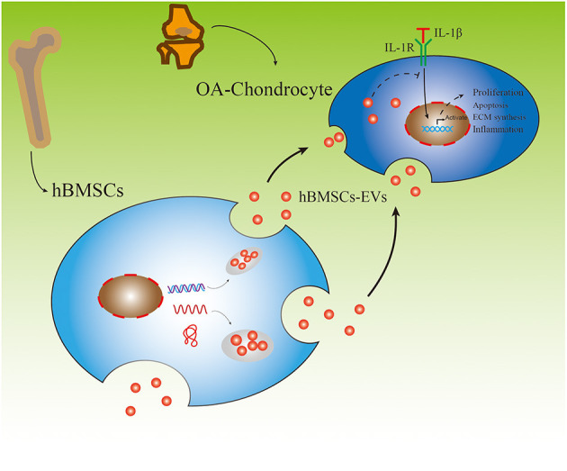
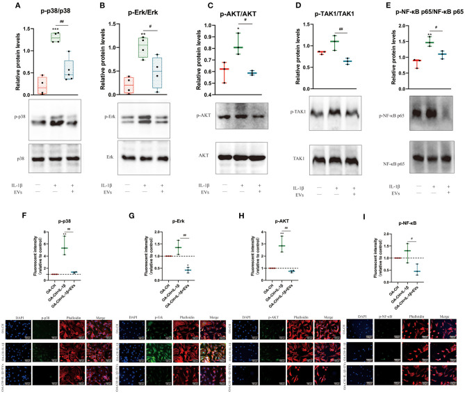
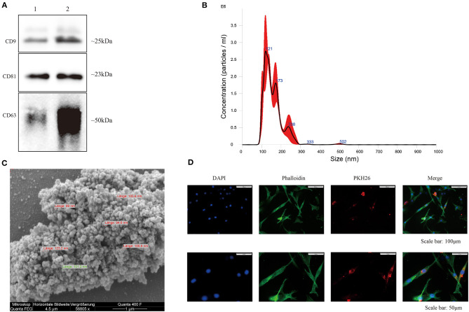
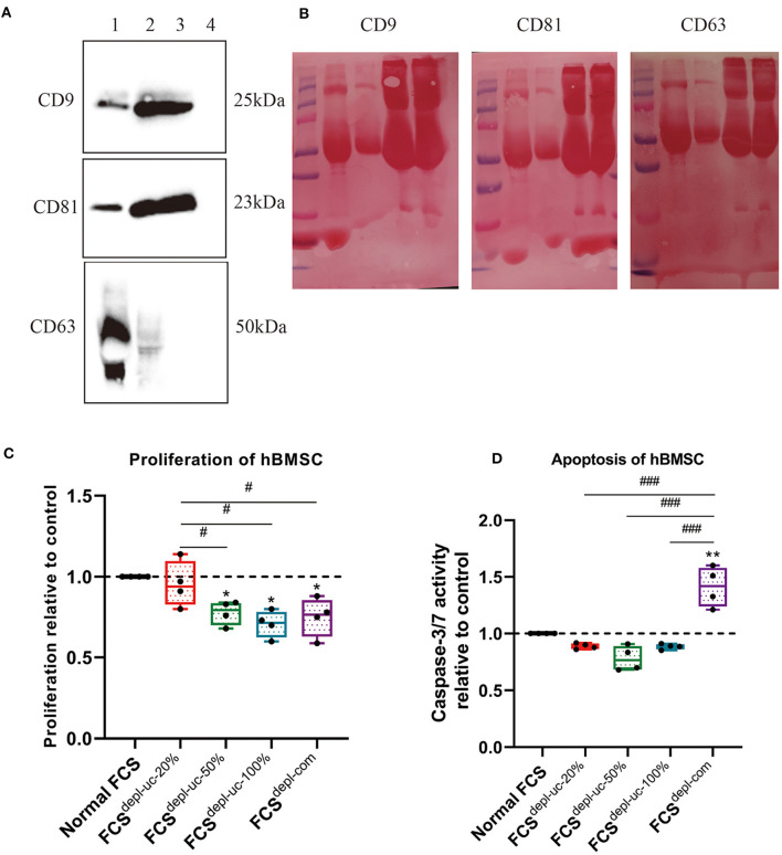
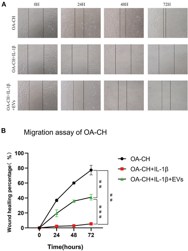
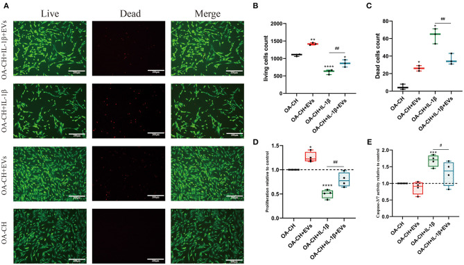
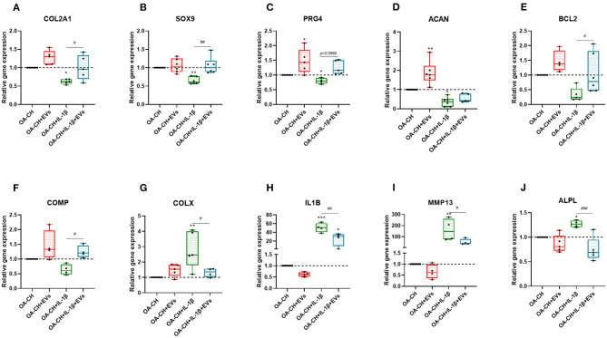
Human bone marrow-derived mesenchymal stromal cells (hBMSCs) provide a promising therapeutic approach in the cell-based therapy of osteoarthritis (OA). However, several disadvantages evolved recently, including immune responses of the host and regulatory hurdles, making it necessary to search for alternative treatment options. Extracellular vesicles (EVs) are released by multiple cell types and tissues into the extracellular microenvironment, acting as message carriers during intercellular communication. Here, we investigate putative protective effects of hBMSC-derived EVs as a cell-free approach, on IL-1β-stimulated chondrocytes obtained from OA-patients. EVs were harvested from the cell culture supernatant of hBMSCs by a sequential ultracentrifugation process. Western blot, scanning electron microscopy (SEM), and nanoparticle tracking analysis (NTA) were performed to characterize the purified particles as EVs. Intracellular incorporation of EVs, derived from PHK26-labeled hBMSCs, was tested by adding the labeled EVs to human OA chondrocytes (OA-CH), followed by fluorescence microscopy. Chondrocytes were pre-stimulated with IL-1β for 24 h, followed by EVs treatment for 24 h. Subsequently, proliferation, apoptosis, and migration (wound healing) were analyzed via BrdU assay, caspase 3/7 assay, and scratch assay, respectively. With qRT-PCR, the relative expression level of anabolic and catabolic genes was determined. Furthermore, immunofluorescence microscopy and western blot were performed to evaluate the protein expression and phosphorylation levels of Erk1/2, PI3K/Akt, p38, TAK1, and NF-κB as components of pro-inflammatory signaling pathways in OA-CH. EVs from hBMSCs (hBMSC-EVs) promote proliferation and reduce apoptosis of OA-CH and IL-1β-stimulated OA-CH. Moreover, hBMSC-EVs attenuate IL-1β-induced reduction of chondrocyte migration. Furthermore, hBMSC-EVs increase gene expression of PRG4, BCL2, and ACAN (aggrecan) and decrease gene expression of MMP13, ALPL, and IL1ß in OA-CH. Notably, COL2A1, SOX9, BCL2, ACAN, and COMP gene expression levels were significantly increased in IL-1β EV groups compared with those IL-1β groups without EVs, whereas the gene expression levels of COLX, IL1B, MMP13, and ALPL were significantly decreased in IL-1β EV groups compared to IL-1β groups without EVs. In addition, the phosphorylation status of Erk1/2, PI3K/Akt, p38, TAK1, and NF-κB signaling molecules, induced by IL-1β, is prevented by hBMSC- EVs. EVs derived from hBMSCs alleviated IL-1β-induced catabolic effects on OA-CH via promoting proliferation and migration and reducing apoptosis, probably via downregulation of IL-1ß-activated pro-inflammatory Erk1/2, PI3K/Akt, p38, TAK1, and NF-κB signaling pathways. EVs released from BMSCs may be considered as promising cell-free intervention strategy in cartilage regenerative medicine, avoiding several adverse effects of cell-based regenerative approaches.
人骨髓间充质基质细胞(hBMSCs)为骨关节炎(OA)的细胞疗法提供了一种很有前景的治疗方法。然而,最近出现了一些缺点,包括宿主的免疫反应和监管障碍,这使得有必要寻找替代治疗方案。细胞外囊泡(EVs)由多种细胞类型和组织释放到细胞外微环境中,在细胞间通讯中充当信息载体。在此,我们研究hBMSC来源的EVs作为一种无细胞方法,对从OA患者获取的经白细胞介素-1β(IL-1β)刺激的软骨细胞的假定保护作用。通过连续超速离心过程从hBMSCs的细胞培养上清液中收获EVs。进行蛋白质印迹、扫描电子显微镜(SEM)和纳米颗粒跟踪分析(NTA),以将纯化的颗粒表征为EVs。通过将标记的EVs添加到人OA软骨细胞(OA-CH)中,然后进行荧光显微镜检查,来测试源自用吡喃鎓花青26(PHK26)标记的hBMSCs的EVs的细胞内摄取情况。软骨细胞先用IL-1β预刺激24小时,然后用EVs处理24小时。随后,分别通过BrdU测定、半胱天冬酶3/7测定和划痕试验分析增殖、凋亡和迁移(伤口愈合)情况。通过定量逆转录聚合酶链反应(qRT-PCR)测定合成代谢和分解代谢基因的相对表达水平。此外,进行免疫荧光显微镜检查和蛋白质印迹,以评估OA-CH中作为促炎信号通路组成部分的细胞外信号调节激酶1/2(Erk1/2)、磷脂酰肌醇-3-激酶/蛋白激酶B(PI3K/Akt)、p38丝裂原活化蛋白激酶(p38)、转化生长因子-β激活激酶1(TAK1)和核因子κB(NF-κB)蛋白表达和磷酸化水平。hBMSCs来源的EVs(hBMSC-EVs)促进OA-CH和经IL-1β刺激的OA-CH的增殖并减少其凋亡。此外,hBMSC-EVs减轻IL-1β诱导的软骨细胞迁移减少。此外,hBMSC-EVs增加OA-CH中富含亮氨酸的蛋白聚糖4(PRG4)、B细胞淋巴瘤2(BCL2)和聚集蛋白聚糖(ACAN)的基因表达,并降低基质金属蛋白酶13(MMP13)、碱性磷酸酶(ALPL)和IL1β的基因表达。值得注意的是,与没有EVs的IL-1β组相比,IL-1β EV组中Ⅱ型胶原α1链(COL2A1)、SRY-box转录因子9(SOX9)、BCL2、ACAN和软骨寡聚基质蛋白(COMP)的基因表达水平显著增加,而与没有EVs的IL-1β组相比,IL-1β EV组中Ⅹ型胶原(COLX)、IL1B、MMP13和ALPL的基因表达水平显著降低。此外,hBMSC-EVs可阻止由IL-1β诱导的Erk1/2、PI3K/Akt、p38、TAK1和NF-κB信号分子的磷酸化状态。hBMSCs来源的EVs可能通过促进增殖和迁移以及减少凋亡,可能通过下调IL-1β激活的促炎Erk1/2、PI3K/Akt、p38、TAK1和NF-κB信号通路,减轻IL-1β对OA-CH的分解代谢作用。BMSCs释放的EVs可被视为软骨再生医学中一种有前景的无细胞干预策略,避免了基于细胞的再生方法的几种不良影响。