Department of Toxicology, School of Public Health, Jilin University, No. 1163 Xinmin Street, Changchun, Jilin, 130021, China.
Department of Pathology, Jilin Medical University, Jilin, China.
Stem Cell Res Ther. 2020 May 11;11(1):174. doi: 10.1186/s13287-020-01616-8.
Skin wounding is very common and may be slow to heal. Increasing evidence shows that exosomes derived from mesenchymal stem cells (MSCs) dramatically enhance skin wound healing in a paracrine manner. However, the mechanism underlying this phenomenon has not yet been elucidated. Thus, the objective of the present study was to identify the signaling pathways and paracrine factors by which MSC-derived exosomes promote de novo skin tissue regeneration in response to wound healing.
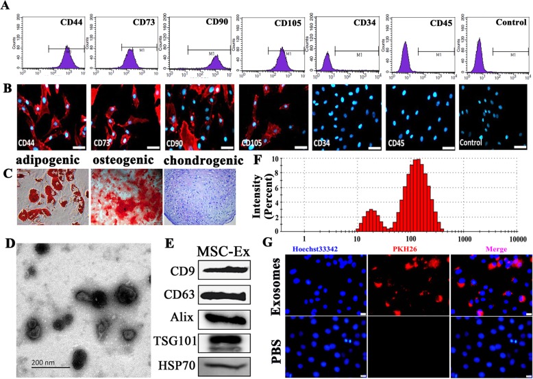
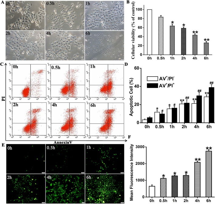
In vitro and in vivo skin wound healing models were created by treating immortalized human keratinocytes (HaCaT) with hydrogen peroxide (HO) and excising full-thickness mouse skin, respectively. Exosomes were extracted from human umbilical cord Wharton's jelly MSCs (hucMSC-Ex) by ultracentrifugation of cell culture supernatant.
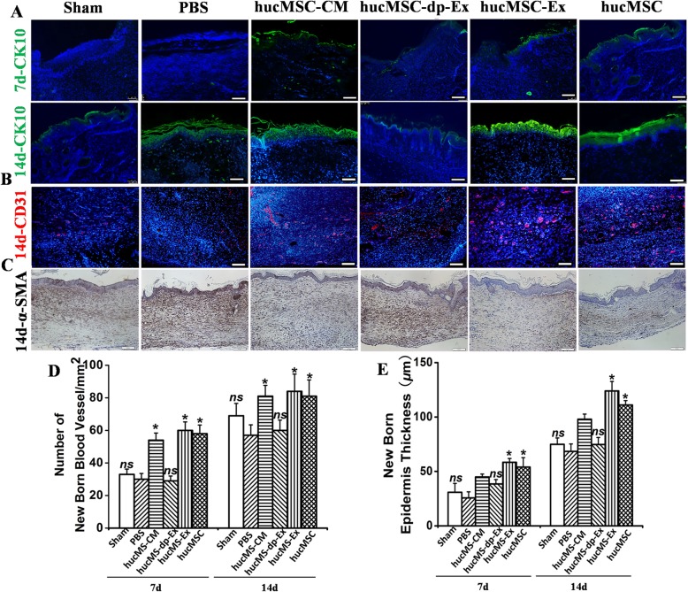
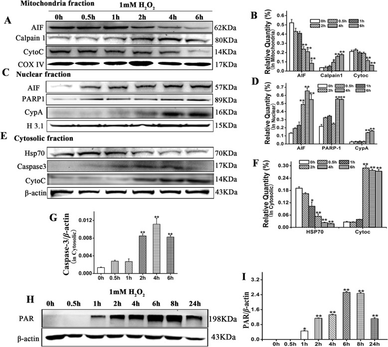
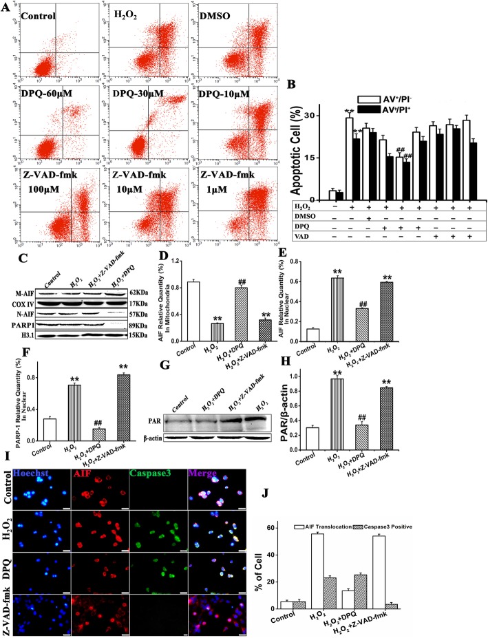
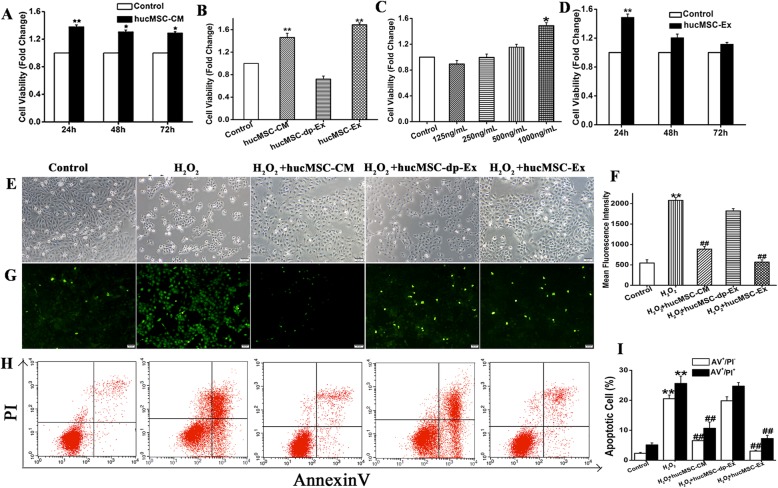
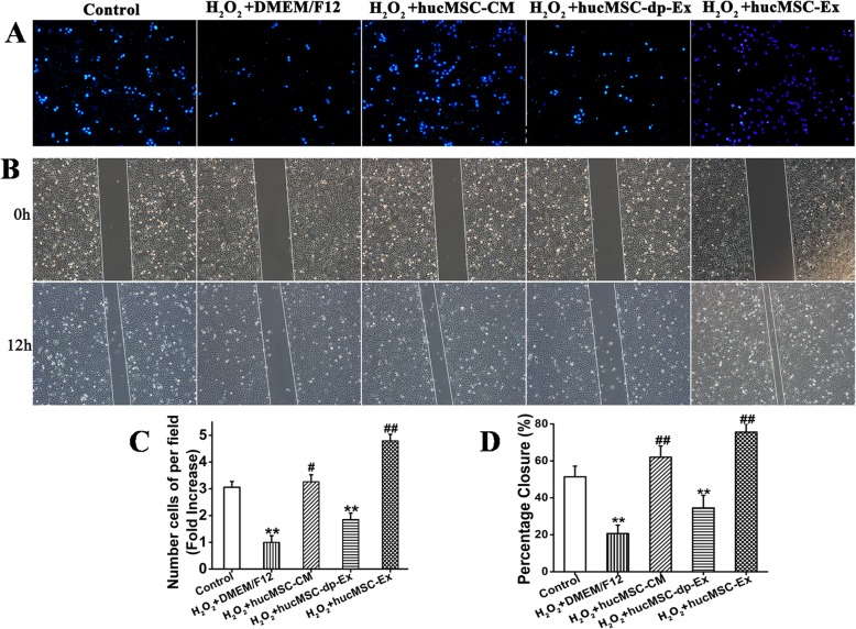
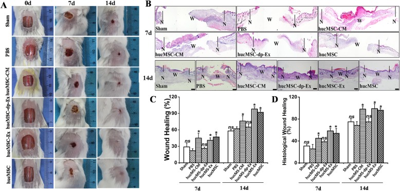
The hucMSC-Ex treatment significantly increased HaCaT cell proliferation and migration in a time- and dose-dependent manner, suppressed HaCaT apoptosis induced with HO by inhibiting nuclear translocation of apoptosis-inducing factor (AIF) and upregulating poly ADP ribose polymerase 1 (PARP-1) and poly (ADP-ribose) (PAR). The animal experiments showed that relative to hucMSCs, hucMSC-Ex attenuated full-thickness skin wounding by enhancing epidermal re-epithelialization and dermal angiogenesis.
These findings indicated that direct administration of hucMSC-Ex may effectively treat cutaneous wounding and could be of great value in clinical settings.
皮肤创伤很常见,且可能愈合缓慢。越来越多的证据表明,间充质干细胞(MSCs)衍生的外泌体通过旁分泌方式显著促进皮肤伤口愈合。然而,这种现象的机制尚未阐明。因此,本研究旨在确定 MSC 衍生的外泌体通过何种信号通路和旁分泌因子促进新的皮肤组织再生以响应伤口愈合。
通过用双氧水(HO)处理永生化人角质细胞(HaCaT)和切除全层小鼠皮肤,分别建立体外和体内皮肤伤口愈合模型。通过超离心细胞培养上清液从人脐带华通氏胶 MSC(hucMSC-Ex)中提取外泌体。
hucMSC-Ex 处理以时间和剂量依赖的方式显著增加 HaCaT 细胞的增殖和迁移,通过抑制凋亡诱导因子(AIF)的核易位和上调多聚 ADP 核糖聚合酶 1(PARP-1)和多聚(ADP-核糖)(PAR)来抑制 HO 诱导的 HaCaT 细胞凋亡。动物实验表明,与 hucMSCs 相比,hucMSC-Ex 通过增强表皮再上皮化和真皮血管生成来减轻全层皮肤创伤。
这些发现表明,直接给予 hucMSC-Ex 可能有效治疗皮肤创伤,在临床环境中具有重要价值。