• 文献检索
  • 文档翻译
  • 深度研究
  • 学术资讯
  • Suppr Zotero 插件Zotero 插件
  • 邀请有礼
  • 套餐&价格
  • 历史记录
应用&插件
Suppr Zotero 插件Zotero 插件浏览器插件Mac 客户端Windows 客户端微信小程序
定价
高级版会员购买积分包购买API积分包
服务
文献检索文档翻译深度研究API 文档MCP 服务
关于我们
关于 Suppr公司介绍联系我们用户协议隐私条款
关注我们

Suppr 超能文献

核心技术专利:CN118964589B侵权必究
粤ICP备2023148730 号-1Suppr @ 2026

文献检索

告别复杂PubMed语法,用中文像聊天一样搜索,搜遍4000万医学文献。AI智能推荐,让科研检索更轻松。

立即免费搜索

文件翻译

保留排版,准确专业,支持PDF/Word/PPT等文件格式,支持 12+语言互译。

免费翻译文档

深度研究

AI帮你快速写综述,25分钟生成高质量综述,智能提取关键信息,辅助科研写作。

立即免费体验

COVID-19 康复患者中,无临床心脏疾病表现但细胞外容积分数升高和整体纵向应变降低。

Elevated Extracellular Volume Fraction and Reduced Global Longitudinal Strains in Participants Recovered from COVID-19 without Clinical Cardiac Findings.

机构信息

From the Departments of Radiology (X.L., T.W., Y.Q., B.L., Y.Y.) and Cardiology (R.Z.), The First Affiliated Hospital of Anhui Medical University, No. 218 Jixi Rd, Hefei 230022, China; Department of Radiology, No. 2 People's Hospital of Fuyang City, Fuyang, China (H.W.); Anhui Province Clinical Image Quality Control Center, Hefei, China (X.L., H.W., Y.Q., B.L., Y.Y.); Department of Radiology, The First Affiliated Hospital of Nanjing Medical University, Nanjing, China (Y.Z.); and Division of Cardiovascular Medicine, Department of Medicine, and Department of Radiology, Perelman School of Medicine, University of Pennsylvania, Philadelphia, Pa (Y.H.).

出版信息

Radiology. 2021 May;299(2):E230-E240. doi: 10.1148/radiol.2021203998. Epub 2021 Jan 12.

DOI:10.1148/radiol.2021203998
PMID:33434112
原文链接:https://pmc.ncbi.nlm.nih.gov/articles/PMC7808090/
Abstract

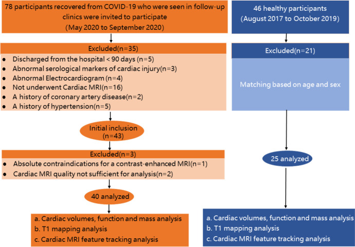
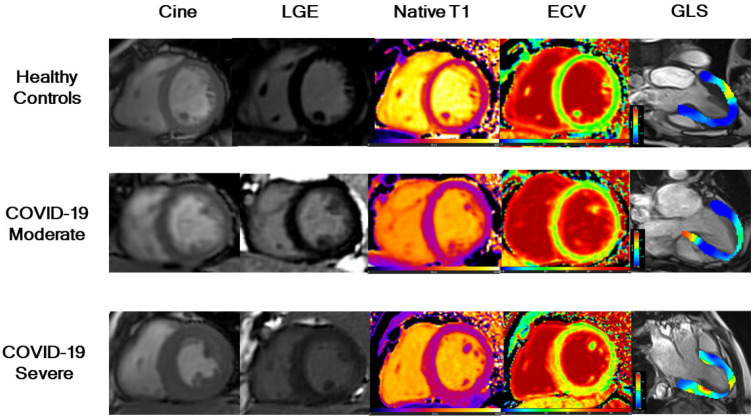
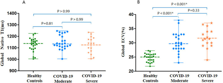
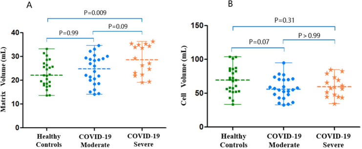
Background It is unknown if there are cardiac abnormalities in persons who have recovered from coronavirus disease 2019 (COVID-19) without cardiac symptoms or in those who have normal biomarkers and normal electrocardiograms. Purpose To evaluate cardiac involvement in participants who had recovered from COVID-19 without clinical evidence of cardiac involvement by using cardiac MRI. Materials and Methods This prospective observational cohort study included 40 participants who had recovered from COVID-19 with moderate ( = 24) or severe ( = 16) pneumonia and who had no cardiovascular medical history, were without cardiac symptoms, had normal electrocardiograms, had normal serologic cardiac enzyme levels, and had been discharged for more than 90 days between May and September 2020. Demographic characteristics were recorded, serum cardiac enzyme levels were measured, and cardiac MRI was performed. Cardiac function, native T1, extracellular volume fraction (ECV), and two-dimensional (2D) strain were quantitatively evaluated and compared with values in control subjects ( = 25). Comparisons among the three groups were performed by using one-way analysis of variance with Bonferroni-corrected post hoc comparisons (for normal distribution) or Kruskal-Wallis tests with post hoc pairwise comparisons (for nonnormal distribution). Results Forty participants (mean age, 54 years ± 12 [standard deviation]; 24 men) were enrolled; participants had a mean time between admission and cardiac MRI of 158 days ± 18 and between discharge and cardiac MRI examination of 124 days ± 17. There were no left or right ventricular size or functional differences between participants who had recovered from COVID-19 and healthy control subjects. Only one (3%) participant had positive late gadolinium enhancement located at the mid inferior wall. Global ECV values were elevated in participants who had recovered from COVID-19 with moderate or severe pneumonia compared with those in healthy control subjects (median ECV, 29.7% vs 31.4% vs 25.0%, respectively; interquartile range, 28.0%-32.9% vs 29.3%-34.0% vs 23.7%-26.0%, respectively; < .001 for both). The 2D global left ventricular longitudinal strain was reduced in both groups of participants (moderate COVID-19 group, -12.5% [interquartile range, -15.5% to -10.7%]; severe COVID-19 group, -12.5% [interquartile range, -15.4% to -8.7%]) compared with the healthy control group (-15.4% [interquartile range, -17.6% to -14.6%]) ( = .002 and = .001, respectively). Conclusion Cardiac MRI myocardial tissue and strain imaging parameters suggest that a proportion of participants who had recovered from COVID-19 had subclinical myocardial abnormalities detectable months after recovery. © RSNA, 2021

摘要

背景 目前尚不清楚在没有心脏症状的情况下从 2019 年冠状病毒病(COVID-19)中康复的患者或在那些具有正常生物标志物和正常心电图的患者中是否存在心脏异常。目的 用心脏 MRI 评估从 COVID-19 中康复且无临床证据表明有心脏受累的患者的心脏受累情况。材料与方法 本前瞻性观察性队列研究纳入了 40 名从 COVID-19 中康复的患者,其中中度(=24 例)或重度(=16 例)肺炎,无心血管病史,无心脏症状,心电图正常,血清心脏酶水平正常,且在 2020 年 5 月至 9 月间出院超过 90 天。记录患者的人口统计学特征,测量血清心脏酶水平,并进行心脏 MRI。对心功能、原生 T1、细胞外容积分数(ECV)和二维(2D)应变进行定量评估,并与对照组(=25 例)的值进行比较。采用单因素方差分析比较三组间的差异,并用 Bonferroni 校正后进行组间两两比较(正态分布)或 Kruskal-Wallis 检验后进行组间两两比较(非正态分布)。结果 共纳入 40 名患者(平均年龄,54 岁±12[标准差];24 名男性);患者入院至心脏 MRI 的平均时间为 158 天±18,出院至心脏 MRI 检查的平均时间为 124 天±17。与健康对照组相比,从 COVID-19 中康复的患者左、右心室大小或功能无差异。仅有 1 名(3%)患者在中下部壁有阳性延迟钆增强。与健康对照组相比,中重度肺炎患者从 COVID-19 中康复后 ECV 值整体升高(中位数 ECV,分别为 29.7%、31.4%和 25.0%;四分位间距,分别为 28.0%-32.9%、29.3%-34.0%和 23.7%-26.0%;均<.001)。两组患者的 2D 整体左心室纵向应变均降低(中度 COVID-19 组,-12.5%[四分位间距,-15.5%至-10.7%];重度 COVID-19 组,-12.5%[四分位间距,-15.4%至-8.7%]),与健康对照组相比[-15.4%(四分位间距,-17.6%至-14.6%)](=.002 和 =.001)。结论 心脏 MRI 心肌组织和应变成像参数提示,一部分从 COVID-19 中康复的患者在康复后数月可检测到亚临床心肌异常。

https://cdn.ncbi.nlm.nih.gov/pmc/blobs/ac49/7808090/8d051820cd04/radiol.2021203998.fig6.jpg
https://cdn.ncbi.nlm.nih.gov/pmc/blobs/ac49/7808090/b9cd8c5ae599/radiol.2021203998.fig1.jpg
https://cdn.ncbi.nlm.nih.gov/pmc/blobs/ac49/7808090/63ef5fc47907/radiol.2021203998.fig2.jpg
https://cdn.ncbi.nlm.nih.gov/pmc/blobs/ac49/7808090/8305b9dcc313/radiol.2021203998.fig3.jpg
https://cdn.ncbi.nlm.nih.gov/pmc/blobs/ac49/7808090/fa9951ee555f/radiol.2021203998.fig4.jpg
https://cdn.ncbi.nlm.nih.gov/pmc/blobs/ac49/7808090/9ab6b39f13f5/radiol.2021203998.fig5.jpg
https://cdn.ncbi.nlm.nih.gov/pmc/blobs/ac49/7808090/8d051820cd04/radiol.2021203998.fig6.jpg

相似文献

1
Elevated Extracellular Volume Fraction and Reduced Global Longitudinal Strains in Participants Recovered from COVID-19 without Clinical Cardiac Findings.COVID-19 康复患者中,无临床心脏疾病表现但细胞外容积分数升高和整体纵向应变降低。
Radiology. 2021 May;299(2):E230-E240. doi: 10.1148/radiol.2021203998. Epub 2021 Jan 12.
2
Cardiac MRI in Patients with Prolonged Cardiorespiratory Symptoms after Mild to Moderate COVID-19.轻度至中度 COVID-19 后出现长时间心肺症状患者的心脏 MRI。
Radiology. 2021 Dec;301(3):E419-E425. doi: 10.1148/radiol.2021211162. Epub 2021 Aug 10.
3
Outcomes of Cardiovascular Magnetic Resonance Imaging in Patients Recently Recovered From Coronavirus Disease 2019 (COVID-19).COVID-19 患者近期康复后的心血管磁共振成像结果。
JAMA Cardiol. 2020 Nov 1;5(11):1265-1273. doi: 10.1001/jamacardio.2020.3557.
4
Observation of Myocardial Involvement in Patients Recovered From COVID-19 by Using Cardiac Magnetic Resonance Imaging, In a Tertiary Care Hospital, Bangladesh.使用心脏磁共振成像观察孟加拉国一家三级保健医院中 COVID-19 康复患者的心肌受累情况。
Mymensingh Med J. 2022 Oct;31(4):1108-1114.
5
T2-mapping increase is the prevalent imaging biomarker of myocardial involvement in active COVID-19: a Cardiovascular Magnetic Resonance study.T2 映射增加是活动性 COVID-19 中心肌受累的主要影像学生物标志物:一项心血管磁共振研究。
J Cardiovasc Magn Reson. 2021 Jun 10;23(1):68. doi: 10.1186/s12968-021-00764-x.
6
Combined Cardiac Fluorodeoxyglucose-Positron Emission Tomography/Magnetic Resonance Imaging Assessment of Myocardial Injury in Patients Who Recently Recovered From COVID-19.近期从 COVID-19 中康复患者的心脏氟脱氧葡萄糖正电子发射断层扫描/磁共振成像心肌损伤的联合评估。
JAMA Cardiol. 2022 Mar 1;7(3):298-308. doi: 10.1001/jamacardio.2021.5505.
7
Ventricular function and tissue characterization by cardiac magnetic resonance imaging following hospitalization for multisystem inflammatory syndrome in children: a prospective study.住院治疗儿童多系统炎症综合征后心脏磁共振成像的心室功能和组织特征:一项前瞻性研究。
Pediatr Radiol. 2023 Mar;53(3):394-403. doi: 10.1007/s00247-022-05521-5. Epub 2022 Oct 18.
8
Cardiac imaging phenotype in patients with coronavirus disease 2019 (COVID-19): results of the cocarde study.2019 年冠状病毒病(COVID-19)患者的心脏影像表型: cocarde 研究结果。
Int J Cardiovasc Imaging. 2021 Feb;37(2):449-457. doi: 10.1007/s10554-020-02010-4. Epub 2020 Sep 9.
9
Cardiac T1 and T2 Mapping Showed Myocardial Involvement in Recovered COVID-19 Patients Initially Considered Devoid of Cardiac Damage.心脏 T1 和 T2 映射显示,最初认为没有心脏损伤的 COVID-19 康复患者存在心肌受累。
J Magn Reson Imaging. 2021 Aug;54(2):421-428. doi: 10.1002/jmri.27534. Epub 2021 Feb 16.
10
MRI T1 Mapping in Hypertrophic Cardiomyopathy: Evaluation in Patients Without Late Gadolinium Enhancement and Hemodynamic Obstruction.磁共振 T1 Mapping 在肥厚型心肌病中的应用:无延迟钆增强和血流动力学梗阻患者的评估。
Radiology. 2020 Feb;294(2):275-286. doi: 10.1148/radiol.2019190651. Epub 2019 Nov 26.

引用本文的文献

1
The value of left ventricular T1 mapping and left atrial strain for distinguishing myocardial amyloidosis and hypertrophic cardiomyopathy.左心室T1映射和左心房应变在鉴别心肌淀粉样变性和肥厚型心肌病中的价值。
Int J Cardiovasc Imaging. 2025 Jul;41(7):1277-1286. doi: 10.1007/s10554-025-03410-0. Epub 2025 Apr 29.
2
Multiparametric cardiovascular magnetic resonance in patients with myocarditis with consecutive follow-up and a comparison between non-COVID-19 and COVID-19-associated myocarditis.对心肌炎患者进行多参数心血管磁共振成像并连续随访,以及非新型冠状病毒肺炎相关心肌炎与新型冠状病毒肺炎相关心肌炎的比较
Quant Imaging Med Surg. 2025 Jan 2;15(1):486-501. doi: 10.21037/qims-24-1422. Epub 2024 Dec 30.
3

本文引用的文献

1
CMR in the Era of COVID-19: Evaluation of Myocarditis in the Subacute Phase.新冠疫情时代的心脏磁共振成像:亚急性期心肌炎的评估
JACC Cardiovasc Imaging. 2020 Nov;13(11):2340-2342. doi: 10.1016/j.jcmg.2020.06.013. Epub 2020 Jul 3.
2
Cardiac Involvement in Patients Recovered From COVID-2019 Identified Using Magnetic Resonance Imaging.利用磁共振成像技术识别 COVID-19 康复患者的心脏受累情况。
JACC Cardiovasc Imaging. 2020 Nov;13(11):2330-2339. doi: 10.1016/j.jcmg.2020.05.004. Epub 2020 May 12.
3
Outcomes of Cardiovascular Magnetic Resonance Imaging in Patients Recently Recovered From Coronavirus Disease 2019 (COVID-19).
Differences in myocardial involvement between new onset and longstanding systemic lupus erythematosus patients assessed by cardiovascular magnetic resonance.
通过心血管磁共振评估新发病和长期系统性红斑狼疮患者心肌受累情况的差异。
Eur J Radiol Open. 2024 Dec 20;14:100623. doi: 10.1016/j.ejro.2024.100623. eCollection 2025 Jun.
4
Immune Profile and MRI-Detected Cardiac Fibrosis and Edema in Hypertensive and Non-Hypertensive Patients with COVID-19.高血压和非高血压COVID-19患者的免疫特征以及MRI检测到的心脏纤维化和水肿
J Clin Med. 2024 Dec 2;13(23):7317. doi: 10.3390/jcm13237317.
5
Extracellular Volume and Fibrosis Volume of Left Ventricular Myocardium Assessed by Cardiac Magnetic Resonance in Vaccinated and Unvaccinated Patients with a History of SARS-CoV-2 Infection.心脏磁共振评估接种和未接种 SARS-CoV-2 感染史患者左心室心肌细胞外体积和纤维化体积。
Cardiovasc Toxicol. 2024 Dec;24(12):1455-1466. doi: 10.1007/s12012-024-09929-3. Epub 2024 Oct 15.
6
Cardiac remodeling and inflammation detected by magnetic resonance imaging in COVID-19 survivors.新冠病毒感染康复者中通过磁共振成像检测到的心脏重塑与炎症
Int J Cardiol Heart Vasc. 2024 Aug 27;54:101499. doi: 10.1016/j.ijcha.2024.101499. eCollection 2024 Oct.
7
Cardiac Magnetic Resonance Imaging of COVID-19-Associated Cardiac Sequelae: A Systematic Review.新冠病毒相关心脏后遗症的心脏磁共振成像:一项系统综述
Rev Cardiovasc Med. 2022 Nov 30;23(12):389. doi: 10.31083/j.rcm2312389. eCollection 2022 Dec.
8
CMR Findings in COVID-19 Recovered Patients: A Review on Parametric Mapping, Feature-Tracking, and LGE.新冠康复患者的心脏磁共振成像(CMR)研究结果:参数映射、特征追踪和延迟强化成像综述
Rev Cardiovasc Med. 2022 Oct 21;23(11):355. doi: 10.31083/j.rcm2311355. eCollection 2022 Nov.
9
Absence of long-term structural and functional cardiac abnormalities on multimodality imaging in a multi-ethnic group of COVID-19 survivors from the early stage of the pandemic.在疫情早期的一个多民族新冠病毒肺炎幸存者群体中,多模态成像显示无长期心脏结构和功能异常。
Eur Heart J Imaging Methods Pract. 2023 Oct 26;1(2):qyad034. doi: 10.1093/ehjimp/qyad034. eCollection 2023 Sep.
10
Myocardial Transit Time Mapping by CMR: A Novel Indicator of Microcirculatory Dysfunction in Cardiac Amyloidosis.通过心脏磁共振成像进行心肌转运时间映射:心脏淀粉样变性中微循环功能障碍的一种新指标。
J Imaging Inform Med. 2024 Dec;37(6):3049-3056. doi: 10.1007/s10278-024-01179-7. Epub 2024 Jun 28.
COVID-19 患者近期康复后的心血管磁共振成像结果。
JAMA Cardiol. 2020 Nov 1;5(11):1265-1273. doi: 10.1001/jamacardio.2020.3557.
4
Pathogenesis and management of myocardial injury in coronavirus disease 2019.新型冠状病毒 2019 型心肌损伤的发病机制和处理。
Eur J Heart Fail. 2020 Nov;22(11):1994-2006. doi: 10.1002/ejhf.1967. Epub 2020 Aug 22.
5
SARS-CoV-2 Infection and Cardiovascular Disease: COVID-19 Heart.新型冠状病毒感染与心血管疾病:COVID-19 心脏。
Heart Lung Circ. 2020 Jul;29(7):973-987. doi: 10.1016/j.hlc.2020.05.101. Epub 2020 Jun 5.
6
CMR and serology to diagnose COVID-19 infection with primary cardiac involvement.心脏磁共振成像(CMR)和血清学检查用于诊断伴有原发性心脏受累的新型冠状病毒肺炎(COVID-19)感染。
Eur Heart J Cardiovasc Imaging. 2021 Jan 1;22(1):133. doi: 10.1093/ehjci/jeaa169.
7
Cardiac MRI in Children with Multisystem Inflammatory Syndrome Associated with COVID-19.COVID-19 相关多系统炎症综合征患儿的心脏 MRI。
Radiology. 2020 Dec;297(3):E283-E288. doi: 10.1148/radiol.2020202288. Epub 2020 Jun 9.
8
Cardiological society of India position statement on COVID-19 and heart failure.印度心脏病学会关于COVID-19与心力衰竭的立场声明。
Indian Heart J. 2020 Mar-Apr;72(2):75-81. doi: 10.1016/j.ihj.2020.04.012. Epub 2020 May 13.
9
Cardiovascular manifestations and treatment considerations in COVID-19.新型冠状病毒肺炎的心血管表现和治疗注意事项。
Heart. 2020 Aug;106(15):1132-1141. doi: 10.1136/heartjnl-2020-317056. Epub 2020 Apr 30.
10
SARS-CoV-2 and viral sepsis: observations and hypotheses.SARS-CoV-2 与病毒败血症:观察与假说。
Lancet. 2020 May 9;395(10235):1517-1520. doi: 10.1016/S0140-6736(20)30920-X. Epub 2020 Apr 17.