Botticchio Alice, Mourad Firas, Fernández-Carnero Samuel, Arias-Buría José Luis, Santodomingo Bueno Alejandro, Mesa Jiménez Juan, Gobbo Massimiliano
Poliambulatorio Physio Power, 25124 Brescia, Italy.
Department of Clinical Science and Translational Medicine, University of Rome Tor Vergata, 00133 Roma, Italy.
J Clin Med. 2021 Jan 8;10(2):209. doi: 10.3390/jcm10020209.
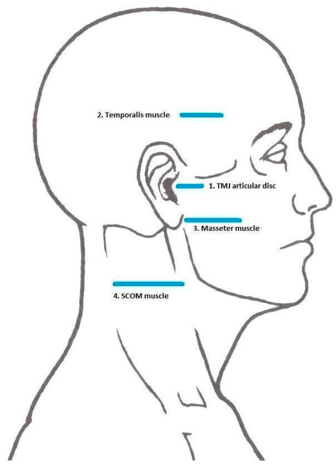
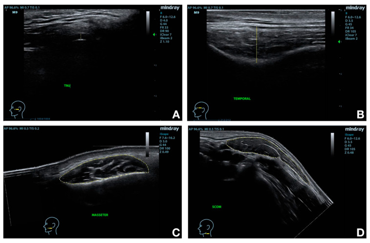
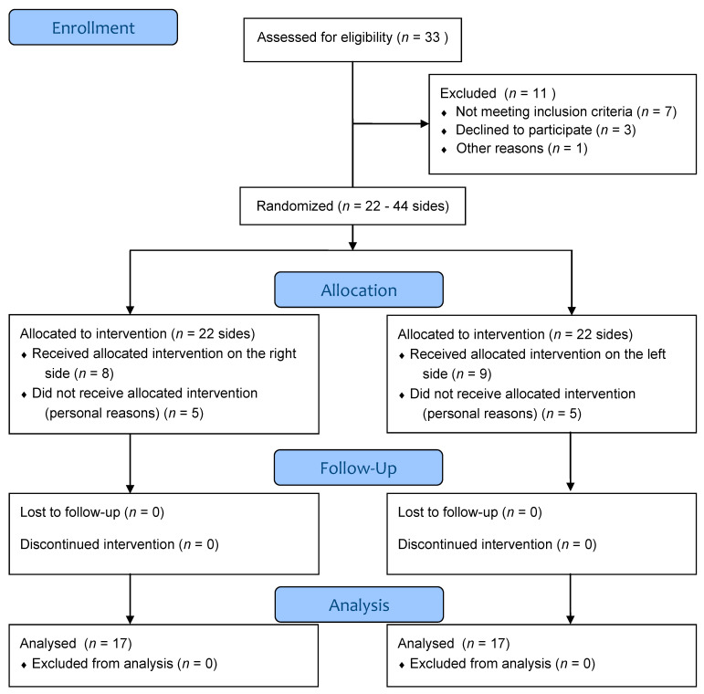
Facial anatomical structures are not easily accessible to manual palpation. The aim of our study is to objectively assess temporomandibular joint and perimandibular muscles dimensions by means of sonographic measurements before and after dry needling (DN) in asymptomatic subjects. Seventeen subjects participated in this before-after study with a within-subject control. After random allocation, one side of the face was used for the intervention and the contralateral as control. DN was performed on the temporal, masseter, and sternocleidomastoid muscles. Each subject was examined bilaterally before, immediately after, and one month after the intervention through Rehabilitative Ultrasound Imaging (RUSI) of the temporomandibular articular disc and the three target muscles. Maximum mouth opening was measured at baseline and at one month. After a single DN session, articular disc thickness significantly decreased; muscles' thicknesses (except for temporal thickness) significantly decreased immediately and at follow-up on the treated side; no significant changes resulted for the control side. The maximum mouth opening increased from 4.77 mm to 4.86 mm. RUSI may be useful to assess the dimensions and thickness of the temporomandibular disc and muscles before and after an intervention. DN influences muscle morphology, and it has a positive influence on mouth opening in the short term.
面部解剖结构不易通过手动触诊触及。我们研究的目的是通过超声测量,客观评估无症状受试者在进行干针疗法(DN)前后颞下颌关节和下颌周围肌肉的尺寸。17名受试者参与了这项前后对照的自身对照研究。随机分组后,一侧面部用于干预,对侧作为对照。在颞肌、咬肌和胸锁乳突肌上进行干针疗法。通过颞下颌关节盘和三块目标肌肉的康复超声成像(RUSI),在干预前、干预后即刻和干预后一个月对每位受试者进行双侧检查。在基线和一个月时测量最大张口度。单次干针治疗后,关节盘厚度显著降低;治疗侧肌肉厚度(颞肌厚度除外)在即刻和随访时均显著降低;对照侧无显著变化。最大张口度从4.77毫米增加到4.86毫米。康复超声成像可能有助于评估干预前后颞下颌关节盘和肌肉的尺寸和厚度。干针疗法会影响肌肉形态,并且在短期内对张口度有积极影响。