• 文献检索
  • 文档翻译
  • 深度研究
  • 学术资讯
  • Suppr Zotero 插件Zotero 插件
  • 邀请有礼
  • 套餐&价格
  • 历史记录
应用&插件
Suppr Zotero 插件Zotero 插件浏览器插件Mac 客户端Windows 客户端微信小程序
定价
高级版会员购买积分包购买API积分包
服务
文献检索文档翻译深度研究API 文档MCP 服务
关于我们
关于 Suppr公司介绍联系我们用户协议隐私条款
关注我们

Suppr 超能文献

核心技术专利:CN118964589B侵权必究
粤ICP备2023148730 号-1Suppr @ 2026

文献检索

告别复杂PubMed语法,用中文像聊天一样搜索,搜遍4000万医学文献。AI智能推荐,让科研检索更轻松。

立即免费搜索

文件翻译

保留排版,准确专业,支持PDF/Word/PPT等文件格式,支持 12+语言互译。

免费翻译文档

深度研究

AI帮你快速写综述,25分钟生成高质量综述,智能提取关键信息,辅助科研写作。

立即免费体验

术后 MRI 精准增强定量作为残余肿瘤影响的指标与胶质母细胞瘤患者的生存相关。

Precise enhancement quantification in post-operative MRI as an indicator of residual tumor impact is associated with survival in patients with glioblastoma.

机构信息

Radiomics Group, Vall d'Hebron Institute of Oncology (VHIO), 117 Natzaret, 08035, Barcelona, Spain.

Department of Radiology, Institut de Diagnòstic Per La Imatge (IDI), Bellvitge University Hospital, Barcelona, Spain.

出版信息

Sci Rep. 2021 Jan 12;11(1):695. doi: 10.1038/s41598-020-79829-3.

DOI:10.1038/s41598-020-79829-3
PMID:33436737
原文链接:https://pmc.ncbi.nlm.nih.gov/articles/PMC7804103/
Abstract

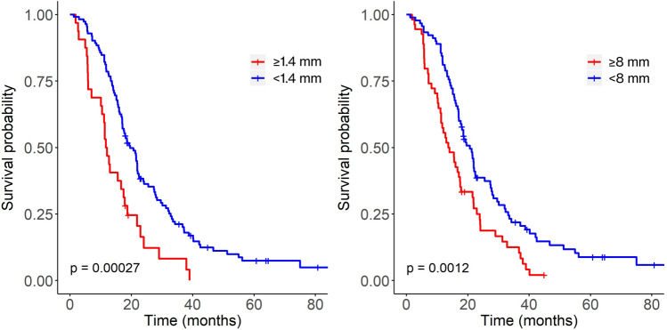
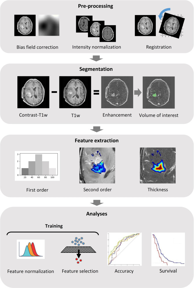
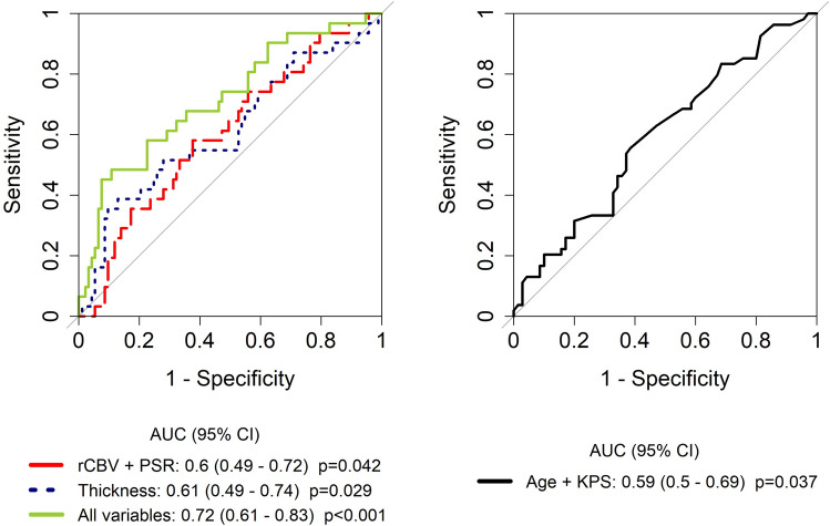
Glioblastoma is the most common primary brain tumor. Standard therapy consists of maximum safe resection combined with adjuvant radiochemotherapy followed by chemotherapy with temozolomide, however prognosis is extremely poor. Assessment of the residual tumor after surgery and patient stratification into prognostic groups (i.e., by tumor volume) is currently hindered by the subjective evaluation of residual enhancement in medical images (magnetic resonance imaging [MRI]). Furthermore, objective evidence defining the optimal time to acquire the images is lacking. We analyzed 144 patients with glioblastoma, objectively quantified the enhancing residual tumor through computational image analysis and assessed the correlation with survival. Pathological enhancement thickness on post-surgical MRI correlated with survival (hazard ratio: 1.98, p < 0.001). The prognostic value of several imaging and clinical variables was analyzed individually and combined (radiomics AUC 0.71, p = 0.07; combined AUC 0.72, p < 0.001). Residual enhancement thickness and radiomics complemented clinical data for prognosis stratification in patients with glioblastoma. Significant results were only obtained for scans performed between 24 and 72 h after surgery, raising the possibility of confounding non-tumor enhancement in very early post-surgery MRI. Regarding the extent of resection, and in agreement with recent studies, the association between the measured tumor remnant and survival supports maximal safe resection whenever possible.

摘要

胶质母细胞瘤是最常见的原发性脑肿瘤。标准疗法包括最大限度地安全切除,辅以辅助放化疗,然后用替莫唑胺进行化疗,但预后极差。目前,手术后残留肿瘤的评估和患者分层为预后组(即根据肿瘤体积)受到医学图像(磁共振成像 [MRI])中残留增强的主观评估的阻碍。此外,缺乏定义获取图像最佳时间的客观证据。我们分析了 144 名胶质母细胞瘤患者,通过计算图像分析客观量化了增强的残留肿瘤,并评估了与生存的相关性。术后 MRI 上的病理性增强厚度与生存相关(风险比:1.98,p<0.001)。单独和联合分析了几种影像学和临床变量的预后价值(放射组学 AUC 为 0.71,p=0.07;联合 AUC 为 0.72,p<0.001)。残留增强厚度和放射组学为胶质母细胞瘤患者的预后分层补充了临床数据。仅在手术后 24 至 72 小时之间进行的扫描中获得了有统计学意义的结果,这增加了在非常早期的术后 MRI 中存在非肿瘤增强的可能性。关于切除范围,与最近的研究一致,测量的肿瘤残余物与生存之间的关联支持尽可能进行最大限度的安全切除。

https://cdn.ncbi.nlm.nih.gov/pmc/blobs/a408/7804103/a4eb6f4d0238/41598_2020_79829_Fig3_HTML.jpg
https://cdn.ncbi.nlm.nih.gov/pmc/blobs/a408/7804103/f47b8a051233/41598_2020_79829_Fig1_HTML.jpg
https://cdn.ncbi.nlm.nih.gov/pmc/blobs/a408/7804103/c06955b4fcf8/41598_2020_79829_Fig2_HTML.jpg
https://cdn.ncbi.nlm.nih.gov/pmc/blobs/a408/7804103/a4eb6f4d0238/41598_2020_79829_Fig3_HTML.jpg
https://cdn.ncbi.nlm.nih.gov/pmc/blobs/a408/7804103/f47b8a051233/41598_2020_79829_Fig1_HTML.jpg
https://cdn.ncbi.nlm.nih.gov/pmc/blobs/a408/7804103/c06955b4fcf8/41598_2020_79829_Fig2_HTML.jpg
https://cdn.ncbi.nlm.nih.gov/pmc/blobs/a408/7804103/a4eb6f4d0238/41598_2020_79829_Fig3_HTML.jpg

相似文献

1
Precise enhancement quantification in post-operative MRI as an indicator of residual tumor impact is associated with survival in patients with glioblastoma.术后 MRI 精准增强定量作为残余肿瘤影响的指标与胶质母细胞瘤患者的生存相关。
Sci Rep. 2021 Jan 12;11(1):695. doi: 10.1038/s41598-020-79829-3.
2
Correlation of the extent of tumor volume resection and patient survival in surgery of glioblastoma multiforme with high-field intraoperative MRI guidance.高场术中磁共振成像引导下多形性胶质母细胞瘤手术中肿瘤体积切除程度与患者生存的相关性。
Neuro Oncol. 2011 Dec;13(12):1339-48. doi: 10.1093/neuonc/nor133. Epub 2011 Sep 12.
3
Maximizing the extent of resection and survival benefit of patients in glioblastoma surgery: high-field iMRI versus conventional and 5-ALA-assisted surgery.最大限度提高胶质母细胞瘤手术患者的切除范围和生存获益:高场强 iMRI 与常规及 5-ALA 辅助手术的比较。
Eur J Surg Oncol. 2014 Mar;40(3):297-304. doi: 10.1016/j.ejso.2013.11.022. Epub 2013 Dec 19.
4
Automated Volumetric Analysis of Postoperative Magnetic Resonance Imaging Predicts Survival in Patients with Glioblastoma.术后磁共振成像的自动容积分析预测胶质母细胞瘤患者的生存。
World Neurosurg. 2019 Jun;126:e1510-e1517. doi: 10.1016/j.wneu.2019.03.142. Epub 2019 Mar 22.
5
Impact on survival of early tumor growth between surgery and radiotherapy in patients with de novo glioblastoma.新诊断胶质母细胞瘤患者手术和放疗之间早期肿瘤生长对生存的影响。
J Neurooncol. 2019 May;142(3):489-497. doi: 10.1007/s11060-019-03120-3. Epub 2019 Feb 19.
6
Complete resection of contrast-enhancing tumor volume is associated with improved survival in recurrent glioblastoma-results from the DIRECTOR trial.对比增强肿瘤体积的完全切除与复发性胶质母细胞瘤患者生存率的提高相关——DIRECTOR试验结果
Neuro Oncol. 2016 Apr;18(4):549-56. doi: 10.1093/neuonc/nov326. Epub 2016 Jan 27.
7
Prognostic Value of Tumor Volume in Glioblastoma Patients: Size Also Matters for Patients with Incomplete Resection.胶质母细胞瘤患者肿瘤体积的预后价值:即使不完全切除,肿瘤大小对患者也很重要。
Ann Surg Oncol. 2018 Feb;25(2):558-564. doi: 10.1245/s10434-017-6253-0. Epub 2017 Nov 20.
8
Magnetic resonance imaging volumetric assessment of the extent of contrast enhancement and resection in oligodendroglial tumors.磁共振成像容积评估寡突胶质细胞瘤对比增强和切除范围。
J Neurosurg. 2012 Jun;116(6):1172-81. doi: 10.3171/2012.2.JNS102032. Epub 2012 Mar 16.
9
Establishing percent resection and residual volume thresholds affecting survival and recurrence for patients with newly diagnosed intracranial glioblastoma.确定影响新诊断颅内胶质母细胞瘤患者生存和复发的百分比切除率和残余体积阈值。
Neuro Oncol. 2014 Jan;16(1):113-22. doi: 10.1093/neuonc/not137. Epub 2013 Nov 26.
10
Accuracy of High-Field Intraoperative MRI in the Detectability of Residual Tumor in Glioma Grade IV Resections.高场强术中磁共振成像在检测四级胶质瘤切除术后残留肿瘤中的准确性
Rofo. 2017 Jun;189(6):519-526. doi: 10.1055/s-0043-106189. Epub 2017 Jun 7.

引用本文的文献

1
Evaluating machine learning models for post-surgery treatment response assessment in glioblastoma multiforme: a comparative study of gray level co-occurrence matrix (GLCM), curvelet, and combined radiomics features selected by multiple algorithms.评估用于多形性胶质母细胞瘤术后治疗反应评估的机器学习模型:灰度共生矩阵(GLCM)、曲波变换以及多种算法选择的联合放射组学特征的比较研究
BMC Med Imaging. 2025 Sep 1;25(1):362. doi: 10.1186/s12880-025-01906-8.
2
Multidisciplinary, Clinical Assessment of Accelerated Deep-Learning MRI Protocols at 1.5 T and 3 T After Intracranial Tumor Surgery and Their Influence on Residual Tumor Perception.颅内肿瘤手术后1.5T和3T加速深度学习MRI协议的多学科临床评估及其对残余肿瘤感知的影响。
Diagnostics (Basel). 2025 Aug 7;15(15):1982. doi: 10.3390/diagnostics15151982.
3

本文引用的文献

1
Automated Volumetric Analysis of Postoperative Magnetic Resonance Imaging Predicts Survival in Patients with Glioblastoma.术后磁共振成像的自动容积分析预测胶质母细胞瘤患者的生存。
World Neurosurg. 2019 Jun;126:e1510-e1517. doi: 10.1016/j.wneu.2019.03.142. Epub 2019 Mar 22.
2
Early response assessment of glioma patients to definitive chemoradiotherapy using chemical exchange saturation transfer imaging at 7 T.7T 下化学交换饱和转移成像在脑胶质瘤患者根治性放化疗早期疗效评估中的应用
J Magn Reson Imaging. 2019 Oct;50(4):1268-1277. doi: 10.1002/jmri.26702. Epub 2019 Mar 12.
3
Gray-level discretization impacts reproducible MRI radiomics texture features.
Comparative Clinical-Imaging and Histogenetic Analysis Between Astrocytoma IDH-Mutant Grade 4 and Glioblastoma IDH-Wildtype-Is There Really a Worse One?4级异柠檬酸脱氢酶(IDH)突变型星形细胞瘤与IDH野生型胶质母细胞瘤的临床影像学与组织发生学比较分析——真的存在更恶性的一种吗?
Diagnostics (Basel). 2025 Feb 11;15(4):438. doi: 10.3390/diagnostics15040438.
4
Radiomics for residual tumour detection and prognosis in newly diagnosed glioblastoma based on postoperative [C] methionine PET and T1c-w MRI.基于术后[C]蛋氨酸 PET 和 T1c-w MRI 的新诊断胶质母细胞瘤中残留肿瘤的检测和预后的影像组学
Sci Rep. 2024 Feb 25;14(1):4576. doi: 10.1038/s41598-024-55092-8.
5
Postoperative Magnetic Resonance Imaging (MRI) Scans for the Surgical Resection of Cranial Glial Tumors According to National Institute of Health and Care Excellence (NICE) Guidelines: A Single-Center Experience.根据英国国家卫生与临床优化研究所(NICE)指南进行颅脑胶质瘤手术切除术后的磁共振成像(MRI)扫描:单中心经验
Cureus. 2023 Dec 24;15(12):e51037. doi: 10.7759/cureus.51037. eCollection 2023 Dec.
6
Artificial Intelligence Analysis Using MRI and PET Imaging in Gliomas: A Narrative Review.使用MRI和PET成像的人工智能分析在胶质瘤中的应用:一项叙述性综述
Cancers (Basel). 2024 Jan 18;16(2):407. doi: 10.3390/cancers16020407.
7
Predicting the WHO Grading of Pediatric Brain Tumors Based on Their MRI Appearance: A Retrospective Study.基于MRI表现预测儿童脑肿瘤的世界卫生组织分级:一项回顾性研究。
Cureus. 2023 Oct 19;15(10):e47333. doi: 10.7759/cureus.47333. eCollection 2023 Oct.
8
Segmentation of glioblastomas in early post-operative multi-modal MRI with deep neural networks.基于深度神经网络的多模态 MRI 术后早期胶质母细胞瘤分割。
Sci Rep. 2023 Nov 2;13(1):18897. doi: 10.1038/s41598-023-45456-x.
9
Resection of positive tissue on methionine-PET is associated with improved survival in glioblastomas.甲硫氨酸-PET 阳性组织切除与胶质母细胞瘤患者的生存改善相关。
Brain Behav. 2023 Dec;13(12):e3291. doi: 10.1002/brb3.3291. Epub 2023 Oct 16.
10
Advanced Magnetic Resonance Imaging in the Evaluation of Treated Glioblastoma: A Pictorial Essay.高级磁共振成像在治疗后胶质母细胞瘤评估中的应用:图文综述
Cancers (Basel). 2023 Jul 26;15(15):3790. doi: 10.3390/cancers15153790.
灰度离散化影响可重现的 MRI 放射组学纹理特征。
PLoS One. 2019 Mar 7;14(3):e0213459. doi: 10.1371/journal.pone.0213459. eCollection 2019.
4
Relaxation-compensated amide proton transfer (APT) MRI signal intensity is associated with survival and progression in high-grade glioma patients.弛豫增强酰胺质子转移(APT)MRI 信号强度与高级别胶质瘤患者的生存和进展相关。
Eur Radiol. 2019 Sep;29(9):4957-4967. doi: 10.1007/s00330-019-06066-2. Epub 2019 Feb 26.
5
CBTRUS Statistical Report: Primary Brain and Other Central Nervous System Tumors Diagnosed in the United States in 2011-2015.CBTRUS统计报告:2011 - 2015年美国原发性脑肿瘤及其他中枢神经系统肿瘤诊断情况
Neuro Oncol. 2018 Oct 1;20(suppl_4):iv1-iv86. doi: 10.1093/neuonc/noy131.
6
Radiomics based on multicontrast MRI can precisely differentiate among glioma subtypes and predict tumour-proliferative behaviour.多对比度 MRI 影像组学可精准区分胶质瘤亚型并预测肿瘤增殖行为。
Eur Radiol. 2019 Apr;29(4):1986-1996. doi: 10.1007/s00330-018-5704-8. Epub 2018 Oct 12.
7
Diagnostic accuracy of MRI texture analysis for grading gliomas.MRI 纹理分析在胶质瘤分级中的诊断准确性。
J Neurooncol. 2018 Dec;140(3):583-589. doi: 10.1007/s11060-018-2984-4. Epub 2018 Aug 25.
8
Evaluation of Glioblastoma Response to Therapy With Chemical Exchange Saturation Transfer.应用化学交换饱和转移评估脑胶质母细胞瘤的治疗反应
Int J Radiat Oncol Biol Phys. 2018 Jul 1;101(3):713-723. doi: 10.1016/j.ijrobp.2018.03.057. Epub 2018 Apr 4.
9
Multisite Concordance of DSC-MRI Analysis for Brain Tumors: Results of a National Cancer Institute Quantitative Imaging Network Collaborative Project.多中心脑肿瘤 DSC-MRI 分析一致性:美国国立癌症研究所定量成像网络协作项目的结果。
AJNR Am J Neuroradiol. 2018 Jun;39(6):1008-1016. doi: 10.3174/ajnr.A5675. Epub 2018 May 24.
10
Validation of postoperative residual contrast-enhancing tumor volume as an independent prognostic factor for overall survival in newly diagnosed glioblastoma.验证术后残留增强肿瘤体积作为新诊断胶质母细胞瘤患者总生存期的独立预后因素。
Neuro Oncol. 2018 Aug 2;20(9):1240-1250. doi: 10.1093/neuonc/noy053.