• 文献检索
  • 文档翻译
  • 深度研究
  • 学术资讯
  • Suppr Zotero 插件Zotero 插件
  • 邀请有礼
  • 套餐&价格
  • 历史记录
应用&插件
Suppr Zotero 插件Zotero 插件浏览器插件Mac 客户端Windows 客户端微信小程序
定价
高级版会员购买积分包购买API积分包
服务
文献检索文档翻译深度研究API 文档MCP 服务
关于我们
关于 Suppr公司介绍联系我们用户协议隐私条款
关注我们

Suppr 超能文献

核心技术专利:CN118964589B侵权必究
粤ICP备2023148730 号-1Suppr @ 2026

文献检索

告别复杂PubMed语法,用中文像聊天一样搜索,搜遍4000万医学文献。AI智能推荐,让科研检索更轻松。

立即免费搜索

文件翻译

保留排版,准确专业,支持PDF/Word/PPT等文件格式,支持 12+语言互译。

免费翻译文档

深度研究

AI帮你快速写综述,25分钟生成高质量综述,智能提取关键信息,辅助科研写作。

立即免费体验

系统性硬化症中视网膜和脉络膜微血管损伤的相关性。

Correlation of retinal and choroidal microvascular impairment in systemic sclerosis.

机构信息

Department of Ophthalmology, University of Lübeck, Ratzeburger Allee 160, 23562, Lübeck, Germany.

Laboratory for Angiogenesis and Ocular Cell Transplantation, University of Lübeck, Ratzeburger Allee 160, 23562, Lübeck, Germany.

出版信息

Orphanet J Rare Dis. 2021 Jan 13;16(1):27. doi: 10.1186/s13023-020-01649-5.

DOI:10.1186/s13023-020-01649-5
PMID:33441156
原文链接:https://pmc.ncbi.nlm.nih.gov/articles/PMC7807887/
Abstract

PURPOSE

To investigate the correlation between retinal and choroidal microperfusion in patients with systemic sclerosis (SSc) using optical coherence tomography angiography (OCTA).

METHODS

In this cross-sectional study SSc patients without clinical evidence of ocular involvement and healthy, age- and sex-matched volunteers were recruited. Participants underwent specific rheumatological and ophthalmological examinations, including optical coherence tomography (OCT) and OCTA. Retinal and choroidal thicknesses as well as perfusion of the retina and the choroidal sublayers were evaluated.

RESULTS

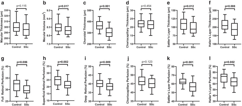
A total of 15 SSc patients (30 eyes) with a median disease duration of 60 months and 15 matched, healthy controls (30 eyes) were recruited. OCT data revealed a significantly lower macular volume, as well as Sattler's layer and Haller's layer thickness in SSc patients compared to controls. In OCTA analysis, the perfusion of both retinal plexus as well as Sattler's and Haller's layer were significantly reduced in the SSc group. Patients with a disease duration of more than 60 months showed a statistically significant positive correlation between retinal and choroidal malperfusion, while those with a shorter disease duration did not.

CONCLUSION

OCTA analysis confirmed impairment of retinal and choroidal microperfusion in SSc patients, supporting the hypothesis of wide spreading vascular injury. In early stages, either the retinal or the choroidal perfusion seems to be involved, while later on, vascular impairment affects both tissues alike. Both, retinal and choroidal examinations should be considered as soon as the diagnosis of SSc is made, to avoid missing out on early alterations.

摘要

目的

应用光学相干断层扫描血管造影术(OCTA)研究系统性硬化症(SSc)患者视网膜和脉络膜微循环的相关性。

方法

本横断面研究纳入了无眼部受累临床表现的 SSc 患者和年龄、性别匹配的健康志愿者。所有参与者均接受了特定的风湿病学和眼科检查,包括光学相干断层扫描(OCT)和 OCTA。评估了视网膜和脉络膜的厚度以及视网膜和脉络膜各层的灌注情况。

结果

共纳入了 15 例 SSc 患者(30 只眼),中位疾病病程为 60 个月,以及 15 名匹配的健康对照者(30 只眼)。OCT 数据显示,与对照组相比,SSc 患者的黄斑容积以及 Sattler 层和 Haller 层厚度显著降低。在 OCTA 分析中,SSc 组视网膜和脉络膜各层的灌注均显著减少。病程超过 60 个月的患者,视网膜和脉络膜灌注不良之间存在统计学上的正相关,而病程较短的患者则无此相关性。

结论

OCTA 分析证实 SSc 患者存在视网膜和脉络膜微循环受损,支持广泛血管损伤的假说。在早期,要么是视网膜灌注,要么是脉络膜灌注受到影响,而在后期,血管损伤则同时影响这两种组织。一旦诊断出 SSc,就应考虑进行视网膜和脉络膜检查,以避免错过早期改变。

https://cdn.ncbi.nlm.nih.gov/pmc/blobs/03a5/7807887/125f5429300c/13023_2020_1649_Fig1_HTML.jpg

相似文献

1
Correlation of retinal and choroidal microvascular impairment in systemic sclerosis.系统性硬化症中视网膜和脉络膜微血管损伤的相关性。
Orphanet J Rare Dis. 2021 Jan 13;16(1):27. doi: 10.1186/s13023-020-01649-5.
2
Evaluation of choroidal substructure perfusion in patients affected by systemic sclerosis: an optical coherence tomography angiography study.评估系统性硬化症患者脉络膜亚结构灌注:光学相干断层扫描血管造影研究。
Scand J Rheumatol. 2020 Mar;49(2):141-145. doi: 10.1080/03009742.2019.1641616. Epub 2019 Sep 17.
3
Choroid, Haller's, and Sattler's layer thickness in intermediate age-related macular degeneration with and without fellow neovascular eyes.伴有和不伴有对侧新生血管性眼的中年年龄相关性黄斑变性中脉络膜、哈勒层和萨特勒层的厚度
Invest Ophthalmol Vis Sci. 2014 Jul 22;55(8):5074-80. doi: 10.1167/iovs.14-14646.
4
Choroidal Haller's and Sattler's layer thickness measurement using 3-dimensional 1060-nm optical coherence tomography.使用三维1060纳米光学相干断层扫描技术测量脉络膜哈勒层和萨特勒层厚度
PLoS One. 2014 Jun 9;9(6):e99690. doi: 10.1371/journal.pone.0099690. eCollection 2014.
5
Elevated serum IGF-1 level enhances retinal and choroidal thickness in untreated acromegaly patients.血清 IGF-1 水平升高可增强未经治疗的肢端肥大症患者的视网膜和脉络膜厚度。
Endocrine. 2018 Mar;59(3):634-642. doi: 10.1007/s12020-017-1511-2. Epub 2018 Jan 10.
6
Decrease in Choroidal Vascularity Index of Haller's layer in diabetic eyes precedes retinopathy.糖尿病眼中 Haller 层脉络膜血管密度指数的下降先于视网膜病变。
BMJ Open Diabetes Res Care. 2020 Sep;8(1). doi: 10.1136/bmjdrc-2020-001295.
7
Peripheral and ocular microvascular alterations in systemic sclerosis: observations from capillaroscopic assessments, perfusion peripheral analysis, and optical coherence tomography angiography.系统性硬化症的外周和眼部微血管改变:从毛细血管镜评估、血流外周分析和光相干断层扫描血管造影观察到的结果。
Rheumatol Int. 2024 Jan;44(1):107-118. doi: 10.1007/s00296-023-05495-z. Epub 2023 Nov 17.
8
Evaluating diurnal changes in choroidal sublayer perfusion using optical coherence tomography angiography.利用光相干断层扫描血管造影评估脉络膜各层灌注的日间变化。
Acta Ophthalmol. 2019 Dec;97(8):e1062-e1068. doi: 10.1111/aos.14140. Epub 2019 May 14.
9
Evaluation of the direct effects on retinal and choroidal microvascularity of systemic scleroderma.系统性硬皮病对视网膜和脉络膜微血管的直接影响评估。
Microvasc Res. 2021 Jul;136:104166. doi: 10.1016/j.mvr.2021.104166. Epub 2021 Apr 9.
10
Evaluating Retinal and Choroidal Perfusion Changes After Ocular Massage of Healthy Eyes Using Optical Coherence Tomography Angiography.评估健康眼行眼部按摩后视网膜和脉络膜血流灌注变化的光学相干断层扫描血管造影研究。
Medicina (Kaunas). 2020 Nov 26;56(12):645. doi: 10.3390/medicina56120645.

引用本文的文献

1
Ophthalmic Posterior Segment OCTA Metrics as Potential Biomarkers for Systemic Involvement in Systemic Sclerosis, Systemic Lupus Erythematosus, and Behçet Disease: A Systematic Review.眼科后段光学相干断层扫描血管造影指标作为系统性硬化症、系统性红斑狼疮和白塞病全身受累的潜在生物标志物:一项系统评价
Open Access Rheumatol. 2025 Apr 26;17:87-100. doi: 10.2147/OARRR.S511810. eCollection 2025.
2
Extrapulmonary manifestations of pulmonary arterial hypertension.肺动脉高压的肺外表现。
Expert Rev Respir Med. 2024 Mar-Apr;18(3-4):189-205. doi: 10.1080/17476348.2024.2361037. Epub 2024 May 30.
3
Ocular Manifestation in Systemic Sclerosis-A Literature Review.

本文引用的文献

1
Analysis of retinal and choroidal microvasculature in systemic sclerosis: an optical coherence tomography angiography study.系统性硬皮病的视网膜和脉络膜微血管分析:一项光学相干断层扫描血管造影研究。
Eye (Lond). 2020 Apr;34(4):763-770. doi: 10.1038/s41433-019-0591-z. Epub 2019 Sep 25.
2
Evaluation of choroidal substructure perfusion in patients affected by systemic sclerosis: an optical coherence tomography angiography study.评估系统性硬化症患者脉络膜亚结构灌注:光学相干断层扫描血管造影研究。
Scand J Rheumatol. 2020 Mar;49(2):141-145. doi: 10.1080/03009742.2019.1641616. Epub 2019 Sep 17.
3
Predictors of disease worsening defined by progression of organ damage in diffuse systemic sclerosis: a European Scleroderma Trials and Research (EUSTAR) analysis.
系统性硬化症的眼部表现——文献综述
Life (Basel). 2024 May 13;14(5):627. doi: 10.3390/life14050627.
4
Retinal Vascular Abnormalities and Clinical Parameters in Systemic Sclerosis.系统性硬化症中的视网膜血管异常与临床参数
J Clin Med. 2024 May 7;13(10):2738. doi: 10.3390/jcm13102738.
5
Macular choroidal thickness, volume, and vascularity index in patients with systemic sclerosis.系统性硬化症患者的黄斑脉络膜厚度、体积和血管指数。
Graefes Arch Clin Exp Ophthalmol. 2024 May;262(5):1475-1487. doi: 10.1007/s00417-023-06342-4. Epub 2023 Dec 22.
6
Retinal microvascular and microstructural alterations in the diagnosis of meibomian gland dysfunction in severely obese population: a new approach.视网膜微血管和微观结构改变在重度肥胖人群睑板腺功能障碍诊断中的应用:一种新方法。
Int J Ophthalmol. 2023 Dec 18;16(12):1977-1985. doi: 10.18240/ijo.2023.12.09. eCollection 2023.
7
Paracentral acute middle maculopathy in systemic sclerosis and subsequent branch retinal artery occlusion.系统性硬化症中的中心旁急性黄斑病变及随后的视网膜分支动脉阻塞
Am J Ophthalmol Case Rep. 2023 Nov 4;32:101955. doi: 10.1016/j.ajoc.2023.101955. eCollection 2023 Dec.
8
Peripheral and ocular microvascular alterations in systemic sclerosis: observations from capillaroscopic assessments, perfusion peripheral analysis, and optical coherence tomography angiography.系统性硬化症的外周和眼部微血管改变:从毛细血管镜评估、血流外周分析和光相干断层扫描血管造影观察到的结果。
Rheumatol Int. 2024 Jan;44(1):107-118. doi: 10.1007/s00296-023-05495-z. Epub 2023 Nov 17.
9
Peripapillary choroidal vascularity index and thickness in patients with systemic sclerosis.系统性硬化症患者的视乳头周围脉络膜血管指数和厚度
Front Med (Lausanne). 2023 Oct 16;10:1273438. doi: 10.3389/fmed.2023.1273438. eCollection 2023.
10
Involvement of the systemic microcirculation in pediatric uveitis.全身性微循环在小儿葡萄膜炎中的作用。
Pediatr Rheumatol Online J. 2023 Oct 2;21(1):109. doi: 10.1186/s12969-023-00896-7.
弥漫性系统性硬化症器官损害进展定义的疾病恶化预测因素:欧洲硬皮病试验和研究(EUSTAR)分析。
Ann Rheum Dis. 2019 Sep;78(9):1242-1248. doi: 10.1136/annrheumdis-2019-215145. Epub 2019 Jun 21.
4
Mapping diurnal variations in choroidal sublayer perfusion in patients with idiopathic epiretinal membrane: an optical coherence tomography angiography study.特发性视网膜前膜患者脉络膜各亚层灌注的昼夜变化图谱:一项光学相干断层扫描血管造影研究
Int J Retina Vitreous. 2019 May 21;5:12. doi: 10.1186/s40942-019-0162-2. eCollection 2019.
5
Evaluating diurnal changes in choroidal sublayer perfusion using optical coherence tomography angiography.利用光相干断层扫描血管造影评估脉络膜各层灌注的日间变化。
Acta Ophthalmol. 2019 Dec;97(8):e1062-e1068. doi: 10.1111/aos.14140. Epub 2019 May 14.
6
Choroidal and retinal thickness in systemic autoimmune and inflammatory diseases: A review.系统性自身免疫和炎症性疾病的脉络膜和视网膜厚度:综述。
Surv Ophthalmol. 2019 Nov-Dec;64(6):757-769. doi: 10.1016/j.survophthal.2019.04.007. Epub 2019 Apr 26.
7
Subfoveal choriocapillaris, Sattler's and Haller's layer thickness predict clinical response to stereotactic radiotherapy in neovascular age-related macular degeneration patients.黄斑中心凹下脉络膜毛细血管、萨特勒层和哈勒层厚度可预测新生血管性年龄相关性黄斑变性患者立体定向放射治疗的临床反应。
J Curr Ophthalmol. 2019 Jan 2;31(1):92-94. doi: 10.1016/j.joco.2018.12.004. eCollection 2019 Mar.
8
Evaluation of retinal microvascular perfusion in systemic sclerosis: a case-control study.系统性硬化症视网膜微血管灌注的评估:一项病例对照研究。
Ann Rheum Dis. 2019 Jun;78(6):857-858. doi: 10.1136/annrheumdis-2018-214541. Epub 2019 Jan 3.
9
Impact of correct anatomical slab segmentation on foveal avascular zone measurements by optical coherence tomography angiography in healthy adults.正确的解剖层面分割对健康成年人光学相干断层扫描血管造影测量黄斑无血管区的影响。
J Curr Ophthalmol. 2018 Mar 20;30(2):156-160. doi: 10.1016/j.joco.2018.02.001. eCollection 2018 Jun.
10
Prevalences of segmentation errors and motion artifacts in OCT-angiography differ among retinal diseases.光学相干断层扫描血管造影(OCT-A)中分割误差和运动伪影的患病率在不同视网膜疾病中存在差异。
Graefes Arch Clin Exp Ophthalmol. 2018 Oct;256(10):1807-1816. doi: 10.1007/s00417-018-4053-2. Epub 2018 Jul 7.