• 文献检索
  • 文档翻译
  • 深度研究
  • 学术资讯
  • Suppr Zotero 插件Zotero 插件
  • 邀请有礼
  • 套餐&价格
  • 历史记录
应用&插件
Suppr Zotero 插件Zotero 插件浏览器插件Mac 客户端Windows 客户端微信小程序
定价
高级版会员购买积分包购买API积分包
服务
文献检索文档翻译深度研究API 文档MCP 服务
关于我们
关于 Suppr公司介绍联系我们用户协议隐私条款
关注我们

Suppr 超能文献

核心技术专利:CN118964589B侵权必究
粤ICP备2023148730 号-1Suppr @ 2026

文献检索

告别复杂PubMed语法,用中文像聊天一样搜索,搜遍4000万医学文献。AI智能推荐,让科研检索更轻松。

立即免费搜索

文件翻译

保留排版,准确专业,支持PDF/Word/PPT等文件格式,支持 12+语言互译。

免费翻译文档

深度研究

AI帮你快速写综述,25分钟生成高质量综述,智能提取关键信息,辅助科研写作。

立即免费体验

使用三维1060纳米光学相干断层扫描技术测量脉络膜哈勒层和萨特勒层厚度

Choroidal Haller's and Sattler's layer thickness measurement using 3-dimensional 1060-nm optical coherence tomography.

作者信息

Esmaeelpour Marieh, Kajic Vedran, Zabihian Behrooz, Othara Richu, Ansari-Shahrezaei Siamak, Kellner Lukas, Krebs Ilse, Nemetz Susanne, Kraus Martin F, Hornegger Joachim, Fujimoto James G, Drexler Wolfgang, Binder Susanne

机构信息

Ludwig Boltzmann Institute of Retinology and Biomicroscopic Laser Surgery, Department of Ophthalmology, Rudolf Foundation Clinic, Vienna, Austria; Center for Medical Physics and Biomedical Engineering, Medical University of Vienna, Vienna, Austria.

Center for Medical Physics and Biomedical Engineering, Medical University of Vienna, Vienna, Austria.

出版信息

PLoS One. 2014 Jun 9;9(6):e99690. doi: 10.1371/journal.pone.0099690. eCollection 2014.

DOI:10.1371/journal.pone.0099690
PMID:24911446
原文链接:https://pmc.ncbi.nlm.nih.gov/articles/PMC4050051/
Abstract

OBJECTIVES

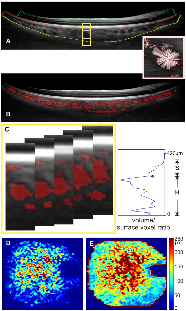
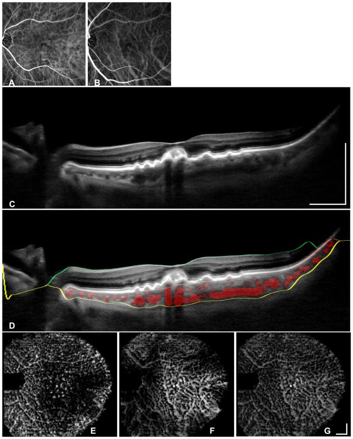
To examine the feasibility of automatically segmented choroidal vessels in three-dimensional (3D) 1060-nmOCT by testing repeatability in healthy and AMD eyes and by mapping Haller's and Sattler's layer thickness in healthy eyes.

METHODS

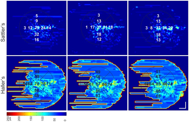
Fifty-five eyes (from 45 healthy subjects and 10 with non-neovascular age-related macular degeneration (AMD) subjects) were imaged by 3D-1060-nmOCT over a 36°x36° field of view. Haller's and Sattler's layer were automatically segmented, mapped and averaged across the Early Treatment Diabetic Retinopathy Study grid. For ten AMD eyes and ten healthy eyes, imaging was repeated within the same session and on another day. Outcomes were the repeatability agreement of Haller's and Sattler's layer thicknesses in healthy and AMD eyes, the validation with ICGA and the statistical analysis of the effect of age and axial eye length (AL) on both healthy choroidal sublayers.

RESULTS

The coefficients of repeatability for Sattler's and Haller's layers were 35% and 21% in healthy eyes and 44% and 31% in AMD eyes, respectively. The mean±SD healthy central submacular field thickness for Sattler's and Haller's was 87±56 µm and 141±50 µm, respectively, with a significant relationship for AL (P<.001).

CONCLUSIONS

Automated Sattler's and Haller's thickness segmentation generates rapid 3D measurements with a repeatability corresponding to reported manual segmentation. Sublayers in healthy eyes thinned significantly with increasing AL. In the presence of the thinned Sattler's layer in AMD, careful measurement interpretation is needed. Automatic choroidal vascular layer mapping may help to explain if pathological choroidal thinning affects medium and large choroidal vasculature in addition to choriocapillaris loss.

摘要

目的

通过检测健康眼和年龄相关性黄斑变性(AMD)眼中的重复性,并绘制健康眼中哈勒层和萨特勒层的厚度,来研究在三维(3D)1060nm光学相干断层扫描(OCT)中自动分割脉络膜血管的可行性。

方法

对55只眼(来自45名健康受试者和10名非新生血管性年龄相关性黄斑变性(AMD)受试者)进行3D-1060nm OCT成像,视野为36°×36°。在糖尿病视网膜病变早期治疗研究网格上自动分割、绘制并平均哈勒层和萨特勒层。对10只AMD眼和10只健康眼在同一检查期间和另一天重复成像。结果包括健康眼和AMD眼中哈勒层和萨特勒层厚度的重复性一致性、与吲哚青绿血管造影(ICGA)的验证以及年龄和眼轴长度(AL)对健康脉络膜两个亚层影响的统计分析。

结果

在健康眼中,萨特勒层和哈勒层的重复性系数分别为35%和21%,在AMD眼中分别为44%和31%。萨特勒层和哈勒层的健康中央黄斑下平均厚度±标准差分别为87±56μm和141±50μm,与AL有显著相关性(P<0.001)。

结论

自动分割萨特勒层和哈勒层厚度可快速进行3D测量,其重复性与报道的手动分割相当。健康眼中的亚层随着AL增加而显著变薄。在AMD患者中,当萨特勒层变薄时,需要仔细解读测量结果。自动脉络膜血管层绘制可能有助于解释病理性脉络膜变薄是否除了影响脉络膜毛细血管丧失外,还会影响中、大脉络膜血管。

https://cdn.ncbi.nlm.nih.gov/pmc/blobs/be43/4050051/334c0c21a77c/pone.0099690.g003.jpg
https://cdn.ncbi.nlm.nih.gov/pmc/blobs/be43/4050051/75b73fc3951c/pone.0099690.g001.jpg
https://cdn.ncbi.nlm.nih.gov/pmc/blobs/be43/4050051/68f15ddddfee/pone.0099690.g002.jpg
https://cdn.ncbi.nlm.nih.gov/pmc/blobs/be43/4050051/334c0c21a77c/pone.0099690.g003.jpg
https://cdn.ncbi.nlm.nih.gov/pmc/blobs/be43/4050051/75b73fc3951c/pone.0099690.g001.jpg
https://cdn.ncbi.nlm.nih.gov/pmc/blobs/be43/4050051/68f15ddddfee/pone.0099690.g002.jpg
https://cdn.ncbi.nlm.nih.gov/pmc/blobs/be43/4050051/334c0c21a77c/pone.0099690.g003.jpg

相似文献

1
Choroidal Haller's and Sattler's layer thickness measurement using 3-dimensional 1060-nm optical coherence tomography.使用三维1060纳米光学相干断层扫描技术测量脉络膜哈勒层和萨特勒层厚度
PLoS One. 2014 Jun 9;9(6):e99690. doi: 10.1371/journal.pone.0099690. eCollection 2014.
2
Choroid, Haller's, and Sattler's layer thickness in intermediate age-related macular degeneration with and without fellow neovascular eyes.伴有和不伴有对侧新生血管性眼的中年年龄相关性黄斑变性中脉络膜、哈勒层和萨特勒层的厚度
Invest Ophthalmol Vis Sci. 2014 Jul 22;55(8):5074-80. doi: 10.1167/iovs.14-14646.
3
Mapping diurnal changes in choroidal, Haller's and Sattler's layer thickness using 3-dimensional 1060-nm optical coherence tomography.使用三维1060纳米光学相干断层扫描技术绘制脉络膜、哈勒层和萨特勒层厚度的昼夜变化图。
Graefes Arch Clin Exp Ophthalmol. 2017 Oct;255(10):1957-1963. doi: 10.1007/s00417-017-3723-9. Epub 2017 Jul 12.
4
Macular Choroidal Small-Vessel Layer, Sattler's Layer and Haller's Layer Thicknesses: The Beijing Eye Study.黄斑脉络膜小血管层、Sattler 层和 Haller 层厚度:北京眼研究。
Sci Rep. 2018 Mar 13;8(1):4411. doi: 10.1038/s41598-018-22745-4.
5
Choroidal thickness in patients with reticular pseudodrusen using 3D 1060-nm OCT maps.利用 3D 1060nm OCT 图谱测量网状假性DR 患者脉络膜厚度。
Invest Ophthalmol Vis Sci. 2014 Apr 25;55(4):2674-81. doi: 10.1167/iovs.13-13338.
6
Choroidal vascularity changes in idiopathic central serous chorioretinopathy after half-fluence photodynamic therapy.特发性中心性浆液性脉络膜视网膜病变半剂量光动力治疗后脉络膜血流变化。
PLoS One. 2018 Aug 27;13(8):e0202930. doi: 10.1371/journal.pone.0202930. eCollection 2018.
7
Decrease in Choroidal Vascularity Index of Haller's layer in diabetic eyes precedes retinopathy.糖尿病眼中 Haller 层脉络膜血管密度指数的下降先于视网膜病变。
BMJ Open Diabetes Res Care. 2020 Sep;8(1). doi: 10.1136/bmjdrc-2020-001295.
8
Choroidal Haller's and Sattler's Layers Thickness in Normal Indian Eyes.正常印度人眼睛脉络膜哈勒层和萨特勒层的厚度
Middle East Afr J Ophthalmol. 2018 Jan-Mar;25(1):19-24. doi: 10.4103/meajo.MEAJO_77_17.
9
Repeatability and reproducibility of choroidal vessel layer measurements in diabetic retinopathy using enhanced depth optical coherence tomography.利用增强深度光学相干断层扫描评估糖尿病视网膜病变脉络膜血管层测量的重复性和可再现性。
Invest Ophthalmol Vis Sci. 2013 Apr 23;54(4):2893-901. doi: 10.1167/iovs.12-11085.
10
En face swept-source optical coherence tomography in neovascular age-related macular degeneration.新生血管性年龄相关性黄斑变性的正面扫频光学相干断层扫描
Br J Ophthalmol. 2015 Sep;99(9):1260-7. doi: 10.1136/bjophthalmol-2014-306422. Epub 2015 Feb 26.

引用本文的文献

1
Three-Dimensional Choroidal Vessel Analysis in Asymmetric Bilateral Age-Related Macular Degeneration: A Comparison of Active Neovascular AMD and Dry AMD Fellow Eyes.不对称双侧年龄相关性黄斑变性的三维脉络膜血管分析:活动性新生血管性年龄相关性黄斑变性与干性年龄相关性黄斑变性对侧眼的比较
Invest Ophthalmol Vis Sci. 2025 Jul 1;66(9):64. doi: 10.1167/iovs.66.9.64.
2
Three-Dimensional Choroidal Vessels Assessment in Diabetic Retinopathy.糖尿病视网膜病变中脉络膜血管的三维评估
Invest Ophthalmol Vis Sci. 2025 Mar 3;66(3):50. doi: 10.1167/iovs.66.3.50.
3
Three-Dimensional Choroidal Vessels Assessment in Age-Related Macular Degeneration.

本文引用的文献

1
Exacerbation of choroidal and retinal pigment epithelial atrophy after anti-vascular endothelial growth factor treatment in neovascular age-related macular degeneration.抗血管内皮生长因子治疗新生血管性年龄相关性黄斑变性后脉络膜和视网膜色素上皮萎缩加重
Retina. 2014 Jul;34(7):1308-15. doi: 10.1097/IAE.0000000000000081.
2
Correlation between subfoveal choroidal thickness and the severity or progression of nonexudative age-related macular degeneration.脉络膜厚度与非渗出性年龄相关性黄斑变性的严重程度或进展的相关性。
Invest Ophthalmol Vis Sci. 2013 Nov 21;54(12):7812-8. doi: 10.1167/iovs.13-12284.
3
Variability of subfoveal choroidal thickness measurements in patients with age-related macular degeneration and central serous chorioretinopathy.
年龄相关性黄斑变性的三维脉络膜血管评估。
Invest Ophthalmol Vis Sci. 2024 Nov 4;65(13):39. doi: 10.1167/iovs.65.13.39.
4
Choroidal imaging biomarkers as predictors of conversion to exudative age-related macular degeneration.脉络膜成像生物标志物作为渗出性年龄相关性黄斑变性转化的预测指标
Graefes Arch Clin Exp Ophthalmol. 2025 Jan;263(1):59-67. doi: 10.1007/s00417-024-06611-w. Epub 2024 Aug 20.
5
Early and Late Treatment Influence on Chorioretinal Microvasculature in Vogt-Koyanagi-Harada Patients Using Optical Coherence Tomography Angiography.采用光学相干断层扫描血管造影术观察早晚期治疗对 Vogt-Koyanagi-Harada 患者脉络膜视网膜微循环的影响。
Transl Vis Sci Technol. 2024 Aug 1;13(8):15. doi: 10.1167/tvst.13.8.15.
6
Automatic segmentation of layers in chorio-retinal complex using Graph-based method for ultra-speed 1.7 MHz wide field swept source FDML optical coherence tomography.基于图论的方法对超快速 1.7MHz 宽场扫频源 FDML 光学相干断层扫描的脉络膜视网膜复合体进行自动分层分割。
Med Biol Eng Comput. 2024 May;62(5):1375-1393. doi: 10.1007/s11517-023-03007-6. Epub 2024 Jan 9.
7
Vessel density changes in choroid, chorio-capillaries, deep and superficial retinal plexues on OCTA in normal ageing and various stages of age-related macular degeneration.在正常衰老和各种年龄相关性黄斑变性阶段,OCTA 中脉络膜、脉络膜毛细血管、深层和浅层视网膜丛的血管密度变化。
Int Ophthalmol. 2023 Oct;43(10):3523-3532. doi: 10.1007/s10792-023-02758-3. Epub 2023 Jun 21.
8
Pathological Neurovascular Unit Mapping onto Multimodal Imaging in Diabetic Macular Edema.病理性神经血管单元在糖尿病性黄斑水肿的多模态成像中的定位。
Medicina (Kaunas). 2023 May 7;59(5):896. doi: 10.3390/medicina59050896.
9
IMI-The Dynamic Choroid: New Insights, Challenges, and Potential Significance for Human Myopia.IMI-动态脉络膜:新的见解、挑战和对人类近视的潜在意义。
Invest Ophthalmol Vis Sci. 2023 May 1;64(6):4. doi: 10.1167/iovs.64.6.4.
10
Within-day changes in luminal, stromal choroidal thickness, and choroidal vascularity index in healthy adults.健康成年人眼内、基质脉络膜厚度和脉络膜血流指数的日内变化。
Indian J Ophthalmol. 2023 Jan;71(1):166-173. doi: 10.4103/ijo.IJO_1268_22.
年龄相关性黄斑变性和中心性浆液性脉络膜视网膜病变患者的中心凹下脉络膜厚度测量值的可变性。
Eye (Lond). 2013 Jul;27(7):809-15. doi: 10.1038/eye.2013.78. Epub 2013 Apr 19.
4
Repeatability and reproducibility of choroidal vessel layer measurements in diabetic retinopathy using enhanced depth optical coherence tomography.利用增强深度光学相干断层扫描评估糖尿病视网膜病变脉络膜血管层测量的重复性和可再现性。
Invest Ophthalmol Vis Sci. 2013 Apr 23;54(4):2893-901. doi: 10.1167/iovs.12-11085.
5
Clinical classification of age-related macular degeneration.年龄相关性黄斑变性的临床分类。
Ophthalmology. 2013 Apr;120(4):844-51. doi: 10.1016/j.ophtha.2012.10.036. Epub 2013 Jan 16.
6
Automated three-dimensional choroidal vessel segmentation of 3D 1060 nm OCT retinal data.对3D 1060纳米光学相干断层扫描(OCT)视网膜数据进行脉络膜血管的自动三维分割。
Biomed Opt Express. 2013 Jan 1;4(1):134-50. doi: 10.1364/BOE.4.000134. Epub 2012 Dec 17.
7
A pilot study of morphometric analysis of choroidal vasculature in vivo, using en face optical coherence tomography.应用光学相干断层扫描对脉络膜血管进行形态计量分析的初步研究。
PLoS One. 2012;7(11):e48631. doi: 10.1371/journal.pone.0048631. Epub 2012 Nov 26.
8
Automated segmentation of the choroid from clinical SD-OCT.从临床 SD-OCT 中自动分割脉络膜。
Invest Ophthalmol Vis Sci. 2012 Nov 1;53(12):7510-9. doi: 10.1167/iovs.12-10311.
9
Motion correction in optical coherence tomography volumes on a per A-scan basis using orthogonal scan patterns.基于正交扫描模式在光学相干断层扫描容积中逐A扫描进行运动校正。
Biomed Opt Express. 2012 Jun 1;3(6):1182-99. doi: 10.1364/BOE.3.001182. Epub 2012 May 3.
10
In vivo human choroidal vascular pattern visualization using high-speed swept-source optical coherence tomography at 1060 nm.在 1060nm 高速扫频源光相干断层扫描下对活体人脉络膜血管形态进行可视化观察。
Invest Ophthalmol Vis Sci. 2012 Apr 30;53(4):2337-48. doi: 10.1167/iovs.11-7823.