Rutka Katarzyna, Garkowski Adam, Karaszewska Katarzyna, Łebkowska Urszula
Department of Radiology, Medical University of Białystok, M. Skłodowskiej-Curie 24A, 15-276 Białystok, Podlaskie, Poland.
Department of Rheumatology and Internal Medicine, Medical University of Białystok, 15-276 Białystok, Podlaskie, Poland.
J Clin Med. 2021 Jan 12;10(2):248. doi: 10.3390/jcm10020248.
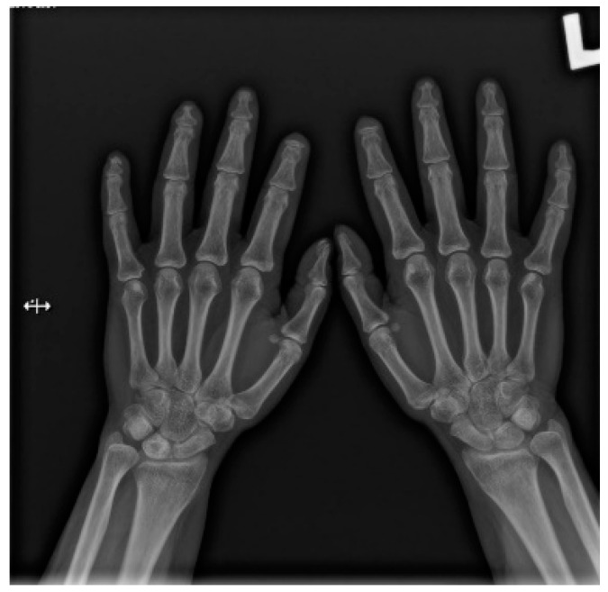
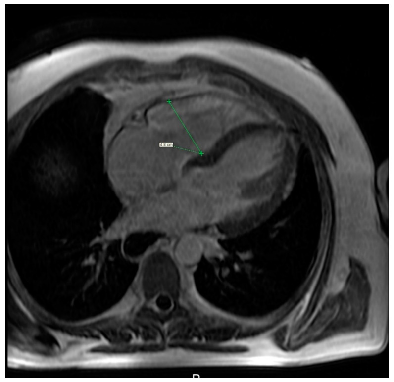
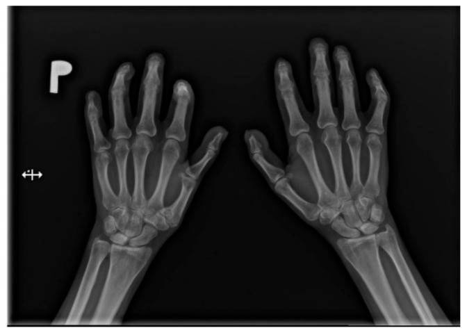
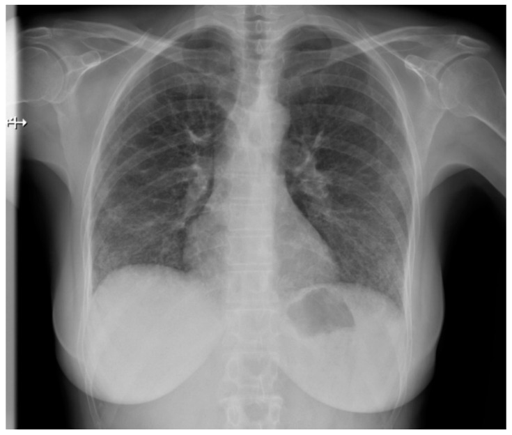
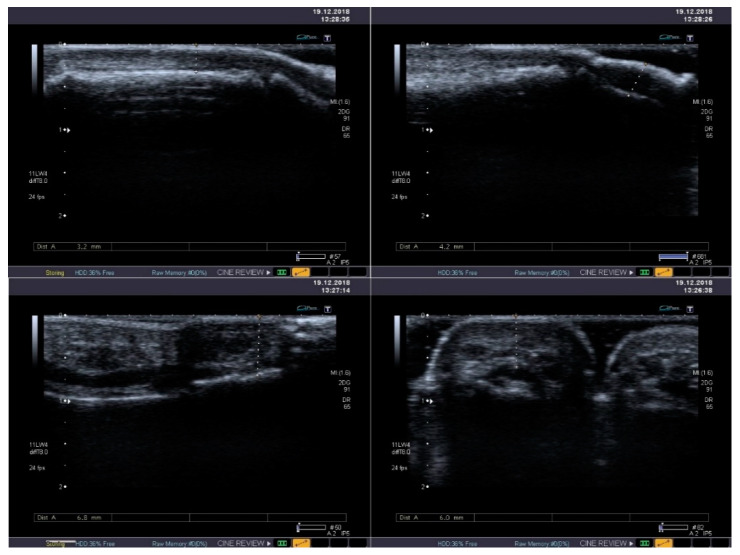
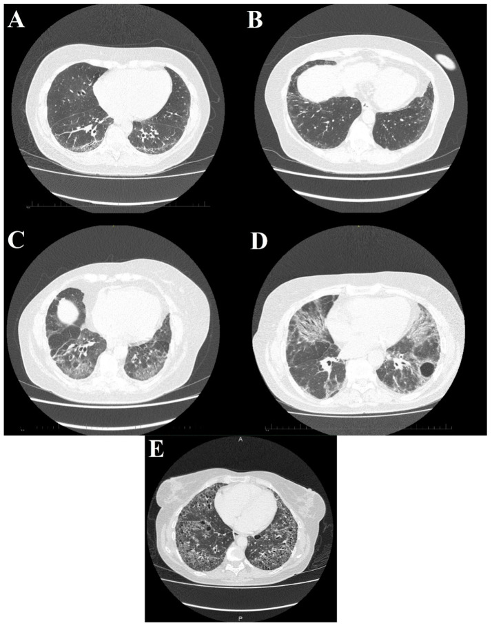
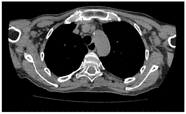
Systemic sclerosis (SSc) is a connective tissue disease characterized by fibrosis in skin and internal organs, progressive vascular obliteration, and the production of autoantibodies. Diagnostic imaging is irreplaceable in both diagnosing and monitoring patients suffering from systemic sclerosis. In addition to routinely used methods, such as comparative X-ray of the hands or a contrast-enhanced examination of the upper gastrointestinal tract or chest, there is an array of less widespread examinations, with an emphasis on magnetic resonance imaging (MRI) and ultrasonography, not only in the evaluation of the musculoskeletal system. This article will review the various imaging modalities available for SSc imaging and assessment, focusing on their utility as tissue-specific diagnosis and treatment monitoring.
系统性硬化症(SSc)是一种结缔组织疾病,其特征为皮肤和内脏器官纤维化、进行性血管闭塞以及自身抗体产生。诊断性影像学在系统性硬化症患者的诊断和监测中具有不可替代的作用。除了常规使用的方法,如手部对比X线检查、上消化道或胸部增强检查外,还有一系列应用较少的检查方法,尤其是磁共振成像(MRI)和超声检查,不仅用于评估肌肉骨骼系统。本文将综述可用于系统性硬化症成像和评估的各种成像方式,重点关注它们在组织特异性诊断和治疗监测方面的效用。