• 文献检索
  • 文档翻译
  • 深度研究
  • 学术资讯
  • Suppr Zotero 插件Zotero 插件
  • 邀请有礼
  • 套餐&价格
  • 历史记录
应用&插件
Suppr Zotero 插件Zotero 插件浏览器插件Mac 客户端Windows 客户端微信小程序
定价
高级版会员购买积分包购买API积分包
服务
文献检索文档翻译深度研究API 文档MCP 服务
关于我们
关于 Suppr公司介绍联系我们用户协议隐私条款
关注我们

Suppr 超能文献

核心技术专利:CN118964589B侵权必究
粤ICP备2023148730 号-1Suppr @ 2026

文献检索

告别复杂PubMed语法,用中文像聊天一样搜索,搜遍4000万医学文献。AI智能推荐,让科研检索更轻松。

立即免费搜索

文件翻译

保留排版,准确专业,支持PDF/Word/PPT等文件格式,支持 12+语言互译。

免费翻译文档

深度研究

AI帮你快速写综述,25分钟生成高质量综述,智能提取关键信息,辅助科研写作。

立即免费体验

使用超灵敏加速器质谱法对 C 胸腺嘧啶核苷标记的间充质干细胞进行体内示踪。

In vivo tracking of C thymidine labeled mesenchymal stem cells using ultra-sensitive accelerator mass spectrometry.

机构信息

Department of Stem Cell Biology, School of Medicine, Konkuk University, 120 Neungdong-Ro, Gwangjin-Gu, 05029, Republic of Korea.

Advanced Analysis Center, Korea Institute of Science and Technology, Hwarang-ro 14-gil 5, Seongbuk-gu, Seoul, 02792, Republic of Korea.

出版信息

Sci Rep. 2021 Jan 14;11(1):1360. doi: 10.1038/s41598-020-80416-9.

DOI:10.1038/s41598-020-80416-9
PMID:33446731
原文链接:https://pmc.ncbi.nlm.nih.gov/articles/PMC7809063/
Abstract

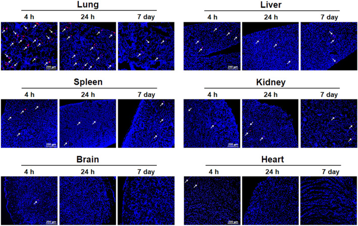
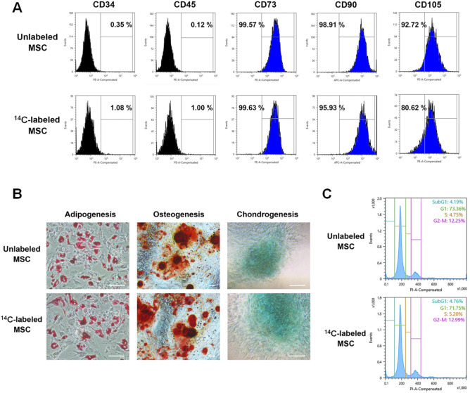
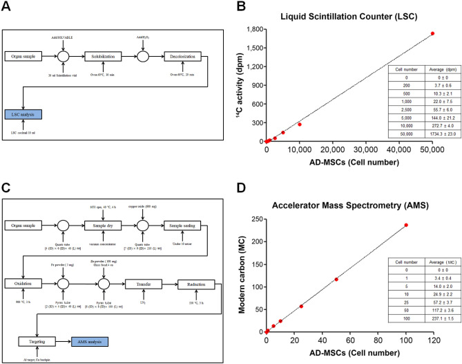
Despite the tremendous advancements made in cell tracking, in vivo imaging and volumetric analysis, it remains difficult to accurately quantify the number of infused cells following stem cell therapy, especially at the single cell level, mainly due to the sensitivity of cells. In this study, we demonstrate the utility of both liquid scintillator counter (LSC) and accelerator mass spectrometry (AMS) in investigating the distribution and quantification of radioisotope labeled adipocyte derived mesenchymal stem cells (AD-MSCs) at the single cell level after intravenous (IV) transplantation. We first show the incorporation of C-thymidine (5 nCi/ml, 24.2 ng/ml) into AD-MSCs without affecting key biological characteristics. These cells were then utilized to track and quantify the distribution of AD-MSCs delivered through the tail vein by AMS, revealing the number of AD-MSCs existing within different organs per mg and per organ at different time points. Notably, the results show that this highly sensitive approach can quantify one cell per mg which effectively means that AD-MSCs can be detected in various tissues at the single cell level. While the significance of these cells is yet to be elucidated, we show that it is possible to accurately depict the pattern of distribution and quantify AD-MSCs in living tissue. This approach can serve to incrementally build profiles of biodistribution for stem cells such as MSCs which is essential for both research and therapeutic purposes.

摘要

尽管在细胞示踪、活体成像和体绘制分析方面取得了巨大进展,但仍然难以准确量化干细胞治疗后输注细胞的数量,特别是在单细胞水平上,这主要是由于细胞的敏感性。在这项研究中,我们展示了液体闪烁计数器 (LSC) 和加速器质谱 (AMS) 在研究放射性同位素标记脂肪间充质干细胞 (AD-MSCs) 在静脉 (IV) 移植后单细胞水平分布和定量的应用。我们首先展示了 C-胸苷(5 nCi/ml,24.2 ng/ml)的掺入对 AD-MSCs 的关键生物学特性没有影响。然后,我们使用这些细胞通过 AMS 追踪和量化通过尾静脉递送的 AD-MSCs 的分布,揭示了不同时间点不同器官每毫克和每个器官中存在的 AD-MSCs 的数量。值得注意的是,结果表明,这种高灵敏度的方法可以定量检测每毫克一个细胞,这有效地意味着 AD-MSCs 可以在各种组织中以单细胞水平检测到。虽然这些细胞的意义尚未阐明,但我们表明可以准确描绘活组织中 AD-MSCs 的分布模式并对其进行定量。这种方法可以为 MSCs 等干细胞建立生物分布概况,这对于研究和治疗目的都是必不可少的。

https://cdn.ncbi.nlm.nih.gov/pmc/blobs/a45a/7809063/ccaf6d9762a2/41598_2020_80416_Fig5_HTML.jpg
https://cdn.ncbi.nlm.nih.gov/pmc/blobs/a45a/7809063/89aeb27d2952/41598_2020_80416_Fig1_HTML.jpg
https://cdn.ncbi.nlm.nih.gov/pmc/blobs/a45a/7809063/ad032670a829/41598_2020_80416_Fig2_HTML.jpg
https://cdn.ncbi.nlm.nih.gov/pmc/blobs/a45a/7809063/293887d253c6/41598_2020_80416_Fig3_HTML.jpg
https://cdn.ncbi.nlm.nih.gov/pmc/blobs/a45a/7809063/13ca2220ddf9/41598_2020_80416_Fig4_HTML.jpg
https://cdn.ncbi.nlm.nih.gov/pmc/blobs/a45a/7809063/ccaf6d9762a2/41598_2020_80416_Fig5_HTML.jpg

相似文献

1
In vivo tracking of C thymidine labeled mesenchymal stem cells using ultra-sensitive accelerator mass spectrometry.使用超灵敏加速器质谱法对 C 胸腺嘧啶核苷标记的间充质干细胞进行体内示踪。
Sci Rep. 2021 Jan 14;11(1):1360. doi: 10.1038/s41598-020-80416-9.
2
In vivo tracking of human placenta derived mesenchymal stem cells in nude mice via ¹⁴C-TdR labeling.通过¹⁴C-胸腺嘧啶核苷标记在裸鼠体内追踪人胎盘来源的间充质干细胞
BMC Biotechnol. 2015 Jun 13;15:55. doi: 10.1186/s12896-015-0174-4.
3
Tracking fusion of human mesenchymal stem cells after transplantation to the heart.追踪人类间充质干细胞移植至心脏后的融合情况。
Stem Cells Transl Med. 2015 Jun;4(6):685-94. doi: 10.5966/sctm.2014-0198. Epub 2015 Apr 6.
4
Noninvasive Tracking and Regenerative Capabilities of Transplanted Human Umbilical Cord-Derived Mesenchymal Stem Cells Labeled with I-III-IV Semiconducting Nanocrystals in Liver-Injured Living Mice.I-III-IV 族半导体纳米晶标记的人脐带间充质干细胞在肝损伤活鼠体内的非侵入性示踪和再生能力。
ACS Appl Mater Interfaces. 2019 Mar 6;11(9):8763-8778. doi: 10.1021/acsami.8b19953. Epub 2019 Feb 21.
5
Homing and Tracking of Iron Oxide Labelled Mesenchymal Stem Cells After Infusion in Traumatic Brain Injury Mice: a Longitudinal In Vivo MRI Study.铁氧化物标记间充质干细胞输注创伤性脑损伤小鼠后的归巢与跟踪:一项纵向体内 MRI 研究。
Stem Cell Rev Rep. 2018 Dec;14(6):888-900. doi: 10.1007/s12015-018-9828-7.
6
Cell tracking of chromium-labeled mesenchymal stem cells using laser ablation inductively coupled plasma imaging mass spectrometry.使用激光烧蚀电感耦合等离子体质谱法对铬标记间充质干细胞进行细胞追踪。
Rapid Commun Mass Spectrom. 2019 Oct 30;33(20):1565-1570. doi: 10.1002/rcm.8505.
7
Human Umbilical Cord Wharton's Jelly-Derived Mesenchymal Stem Cells Labeled with Mn and Gd Co-Doped CuInS-ZnS Nanocrystals for Multimodality Imaging in a Tumor Mice Model.人脐带华通氏胶源间充质干细胞标记 Mn 和 Gd 共掺杂的 CuInS-ZnS 纳米晶体用于肿瘤小鼠模型的多模态成像。
ACS Appl Mater Interfaces. 2020 Jan 22;12(3):3415-3429. doi: 10.1021/acsami.9b19054. Epub 2020 Jan 9.
8
Towards whole-body imaging at the single cell level using ultra-sensitive stem cell labeling with oligo-arginine modified upconversion nanoparticles.利用经过聚精氨酸修饰的上转换纳米粒子对干细胞进行超灵敏标记,实现单细胞水平的全身成像。
Biomaterials. 2012 Jun;33(19):4872-81. doi: 10.1016/j.biomaterials.2012.03.047. Epub 2012 Apr 5.
9
Mesenchymal Stem Cells Delivery in Individuals with Different Pathologies: Multimodal Tracking, Safety and Future Applications.间充质干细胞在不同疾病患者中的递送:多模态追踪、安全性和未来应用。
Int J Mol Sci. 2022 Jan 31;23(3):1682. doi: 10.3390/ijms23031682.
10
MRI tracking of polyethylene glycol-coated superparamagnetic iron oxide-labelled placenta-derived mesenchymal stem cells toward glioblastoma stem-like cells in a mouse model.MRI 示踪聚乙二醇修饰的超顺磁性氧化铁标记胎盘间充质干细胞向荷胶质瘤干细胞小鼠模型中的归巢。
Artif Cells Nanomed Biotechnol. 2018;46(sup3):S448-S459. doi: 10.1080/21691401.2018.1499661. Epub 2018 Sep 8.

引用本文的文献

1
The development and evolution of biological AMS at Livermore: a perspective.利弗莫尔生物加速器质谱法的发展与演变:一个视角
Bioanalysis. 2025 Mar;17(5):345-354. doi: 10.1080/17576180.2025.2460391. Epub 2025 Feb 4.

本文引用的文献

1
Mesenchymal Stromal Cell Therapeutic Delivery: Translational Challenges to Clinical Application.间质基质细胞治疗性递药:向临床应用的转化挑战。
Front Immunol. 2019 Jul 31;10:1645. doi: 10.3389/fimmu.2019.01645. eCollection 2019.
2
The Effect of Hexanoyl Glycol Chitosan on the Proliferation of Human Mesenchymal Stem Cells.己酰基乙二醇壳聚糖对人间充质干细胞增殖的影响
Polymers (Basel). 2018 Jul 30;10(8):839. doi: 10.3390/polym10080839.
3
Macrophage cell tracking PET imaging using mesoporous silica nanoparticles via in vivo bioorthogonal F-18 labeling.
介孔硅纳米粒子通过体内生物正交 F-18 标记进行巨噬细胞示踪 PET 成像。
Biomaterials. 2019 Apr;199:32-39. doi: 10.1016/j.biomaterials.2019.01.043. Epub 2019 Feb 2.
4
Tracking Tumor Colonization in Xenograft Mouse Models Using Accelerator Mass Spectrometry.利用加速器质谱法追踪异种移植小鼠模型中的肿瘤定植。
Sci Rep. 2018 Oct 9;8(1):15013. doi: 10.1038/s41598-018-33368-0.
5
Robust Revascularization in Models of Limb Ischemia Using a Clinically Translatable Human Stem Cell-Derived Endothelial Cell Product.使用一种具有临床转化前景的人源干细胞衍生的内皮细胞产品实现肢体缺血模型中的稳健再血管化。
Mol Ther. 2018 Jul 5;26(7):1669-1684. doi: 10.1016/j.ymthe.2018.03.017. Epub 2018 Mar 28.
6
Tracking of transplanted human umbilical cord-derived mesenchymal stem cells labeled with fluorescent probe in a mouse model of acute lung injury.荧光探针标记的人脐带间充质干细胞移植在急性肺损伤小鼠模型中的示踪研究。
Int J Mol Med. 2018 May;41(5):2527-2534. doi: 10.3892/ijmm.2018.3491. Epub 2018 Feb 13.
7
In vivo stem cell tracking with imageable nanoparticles that bind bioorthogonal chemical receptors on the stem cell surface.利用可成像纳米颗粒对干细胞表面的生物正交化学受体进行体内干细胞示踪。
Biomaterials. 2017 Sep;139:12-29. doi: 10.1016/j.biomaterials.2017.05.050. Epub 2017 Jun 1.
8
Dynamic In Vivo SPECT Imaging of Neural Stem Cells Functionalized with Radiolabeled Nanoparticles for Tracking of Glioblastoma.用于胶质母细胞瘤追踪的放射性标记纳米颗粒功能化神经干细胞的动态体内单光子发射计算机断层扫描成像
J Nucl Med. 2016 Feb;57(2):279-84. doi: 10.2967/jnumed.115.163006. Epub 2015 Nov 12.
9
In vivo tracking of human placenta derived mesenchymal stem cells in nude mice via ¹⁴C-TdR labeling.通过¹⁴C-胸腺嘧啶核苷标记在裸鼠体内追踪人胎盘来源的间充质干细胞
BMC Biotechnol. 2015 Jun 13;15:55. doi: 10.1186/s12896-015-0174-4.
10
In vivo real-time visualization of mesenchymal stem cells tropism for cutaneous regeneration using NIR-II fluorescence imaging.利用近红外二区荧光成像技术在体实时可视化间充质干细胞对皮肤再生的趋向性。
Biomaterials. 2015 Jun;53:265-73. doi: 10.1016/j.biomaterials.2015.02.090. Epub 2015 Mar 16.