Centre for Research and Development in Learning (CRADLE), Nanyang Technological University, CRADLE, 61 Nanyang Drive, ABN-01b-10, Singapore, 637335, Singapore.
Department of Psychiatry, New York University School of Medicine, New York, USA.
Sci Rep. 2021 Jan 14;11(1):1354. doi: 10.1038/s41598-020-80346-6.
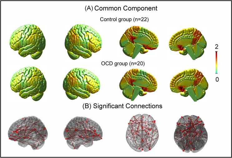
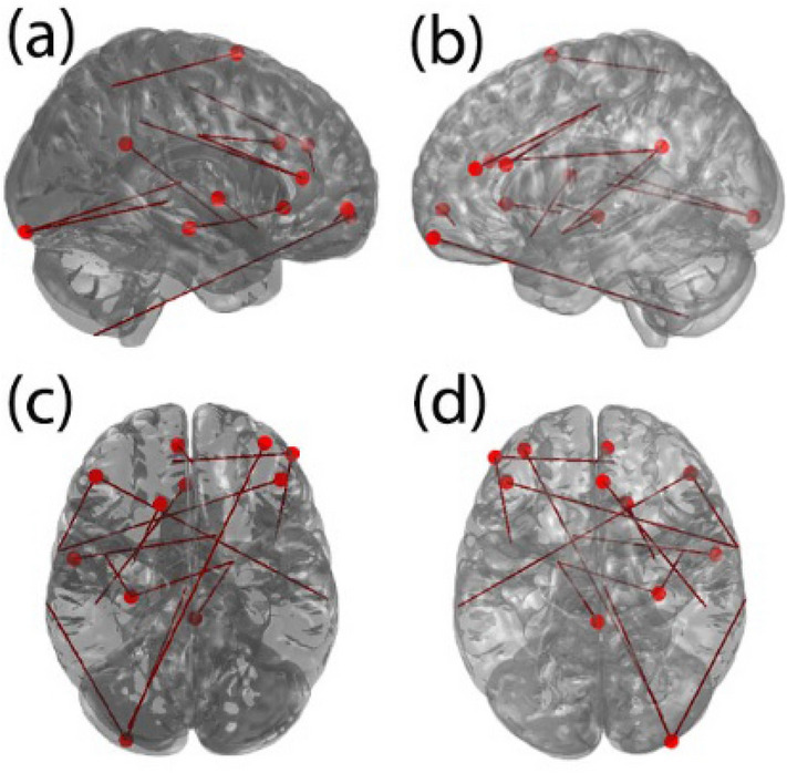
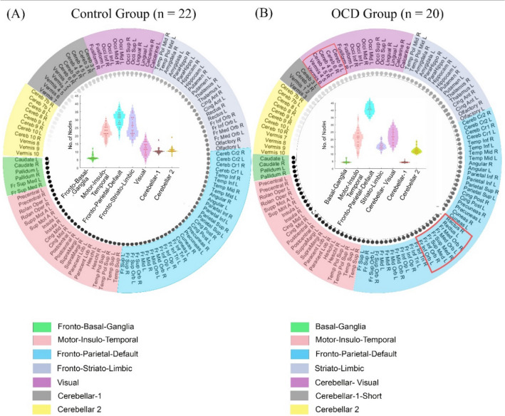
There is significant interest in understanding the pathophysiology of Obsessive-Compulsive Disorder (OCD) using resting-state fMRI (rsfMRI). Previous studies acknowledge abnormalities within and beyond the fronto-striato-limbic circuit in OCD that require further clarifications. However, limited information could be inferred from the conventional way of investigating the functional connectivity differences between OCD and healthy controls. Here, we identified altered brain organization in patients with OCD by applying individual-based approaches to maximize the identification of underlying network-based features specific to the OCD group. rsfMRI of 20 patients with OCD and 22 controls were preprocessed, and individual-fMRI-subspace was derived for each subject within each group. We evaluated group differences in functional connectivity using individual-fMRI-subspace and established its advantage over conventional-fMRI methodology. We applied prediction-based approaches to highlight the group differences by evaluating the differences in functional connections that predicted the clinical scores (namely, the Obsessive-Compulsive Inventory-Revised (OCI-R) and Hamilton Anxiety Rating Scale). Then, we explored the brain network organization of both groups by estimating the subject-specific communities within each group. Lastly, we evaluated associations between the inter-individual variation of nodes in the communities to clinical measures using linear regression. Functional connectivity analysis using individual-fMRI-subspace detected 83 connections that were different between OCD and control groups, compared to none found using conventional-fMRI methodology. Connectome-based prediction analysis did not show significant overlap between the two groups in the functional connections that predicted the clinical scores. This suggests that the functional architecture in patients with OCD may be different compared to controls. Seven communities were found in both groups. Interestingly, within the OCD group but not controls, we observed functional connectivity between cerebellar and visual regions, and lack of connectivity between striato-limbic and frontal areas. Inter-individual variations in the community-size of these two communities were also associated with the OCI-R score (p < .005). Due to our small sample size, we further validated our results by (i) accounting for head motion, (ii) applying global signal regression (GSR) in data processing, and (iii) using an alternate atlas for parcellation. While the main results were consistently observed with accounting for head motion and using another atlas, the key findings were not reproduced with GSR application. The study demonstrated the existence of disconnectedness in fronto-striato-limbic community and connectedness between cerebellar and visual areas in OCD patients, which was also related to the clinical symptomatology of OCD.
人们对使用静息态 fMRI(rsfMRI)来理解强迫症(OCD)的病理生理学非常感兴趣。以前的研究承认 OCD 患者存在额纹状体边缘回路内外的异常,这需要进一步澄清。然而,从传统的 OCD 患者与健康对照组之间功能连接差异的研究方法中,我们只能推断出有限的信息。在这里,我们通过应用个体为基础的方法来最大化识别 OCD 组特有的潜在基于网络的特征,从而确定 OCD 患者大脑组织的改变。对 20 名 OCD 患者和 22 名对照组的 rsfMRI 进行预处理,并为每个组中的每个受试者导出个体 fMRI 子空间。我们使用个体 fMRI 子空间评估功能连接的组间差异,并建立其优于传统 fMRI 方法的优势。我们应用基于预测的方法,通过评估预测临床评分(即强迫症检查表修订版(OCI-R)和汉密尔顿焦虑量表)的功能连接差异来突出组间差异。然后,我们通过估计每个组内的特定于主题的社区来探索两组的大脑网络组织。最后,我们使用线性回归评估社区中节点的个体间变异性与临床指标之间的相关性。与使用传统 fMRI 方法相比,使用个体 fMRI 子空间进行功能连接分析检测到 OCD 和对照组之间有 83 个不同的连接。连接组预测分析未显示两组在预测临床评分的功能连接上有显著重叠。这表明 OCD 患者的功能架构可能与对照组不同。在两组中都发现了七个社区。有趣的是,在 OCD 组中,但在对照组中,我们观察到小脑和视觉区域之间的功能连接,以及纹状体边缘和额叶区域之间缺乏连接。这两个社区的社区大小的个体间变化也与 OCI-R 评分相关(p <.005)。由于我们的样本量较小,因此我们通过以下方式进一步验证了我们的结果:(i)考虑头部运动,(ii)在数据处理中应用全局信号回归(GSR),以及(iii)使用替代图谱进行分割。虽然在考虑头部运动和使用另一个图谱时,主要结果是一致的,但应用 GSR 并未再现关键发现。该研究表明 OCD 患者中存在额纹状体边缘社区的不连接和小脑与视觉区域之间的连接,这与 OCD 的临床症状也有关。