• 文献检索
  • 文档翻译
  • 深度研究
  • 学术资讯
  • Suppr Zotero 插件Zotero 插件
  • 邀请有礼
  • 套餐&价格
  • 历史记录
应用&插件
Suppr Zotero 插件Zotero 插件浏览器插件Mac 客户端Windows 客户端微信小程序
定价
高级版会员购买积分包购买API积分包
服务
文献检索文档翻译深度研究API 文档MCP 服务
关于我们
关于 Suppr公司介绍联系我们用户协议隐私条款
关注我们

Suppr 超能文献

核心技术专利:CN118964589B侵权必究
粤ICP备2023148730 号-1Suppr @ 2026

文献检索

告别复杂PubMed语法,用中文像聊天一样搜索,搜遍4000万医学文献。AI智能推荐,让科研检索更轻松。

立即免费搜索

文件翻译

保留排版,准确专业,支持PDF/Word/PPT等文件格式,支持 12+语言互译。

免费翻译文档

深度研究

AI帮你快速写综述,25分钟生成高质量综述,智能提取关键信息,辅助科研写作。

立即免费体验

低氧诱导因子 1α 促进慢性肺淋巴流量升高条件下淋巴管内皮细胞的增殖。

HIF-1α promotes cellular growth in lymphatic endothelial cells exposed to chronically elevated pulmonary lymph flow.

机构信息

Department of Pediatrics, University of California, San Francisco, 513 Parnassus Avenue, HSE1418, Box 1346, San Francisco, CA, 94143-1346, USA.

Department of Surgery, University of California, Davis, Davis, USA.

出版信息

Sci Rep. 2021 Jan 14;11(1):1468. doi: 10.1038/s41598-020-80882-1.

DOI:10.1038/s41598-020-80882-1
PMID:33446832
原文链接:https://pmc.ncbi.nlm.nih.gov/articles/PMC7809484/
Abstract

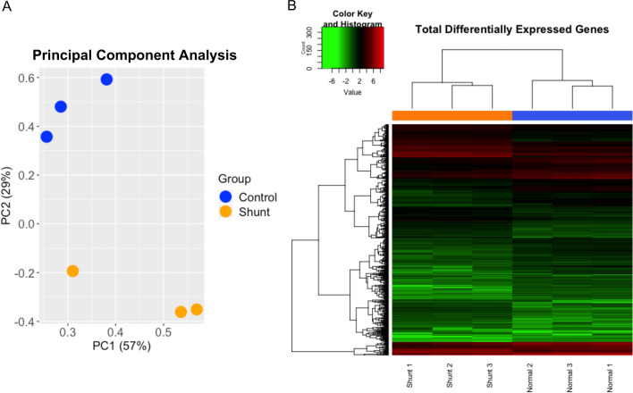
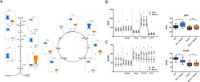
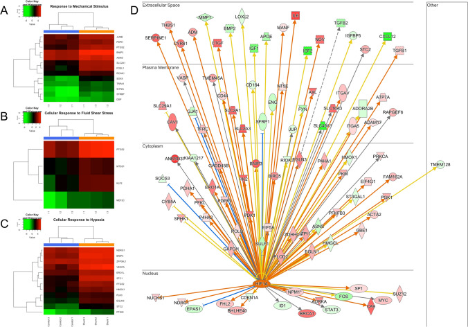
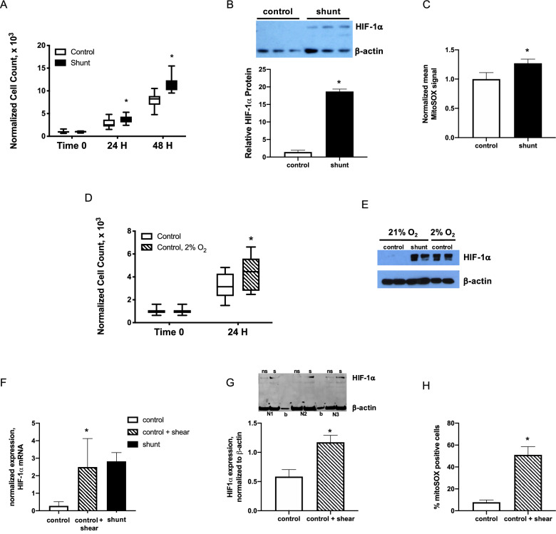
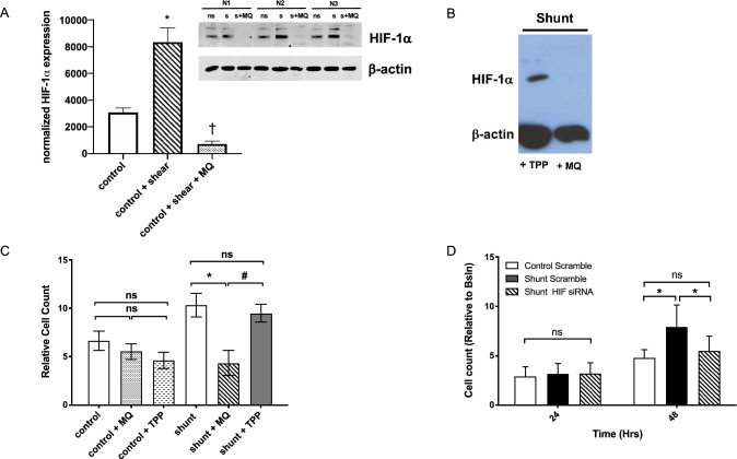
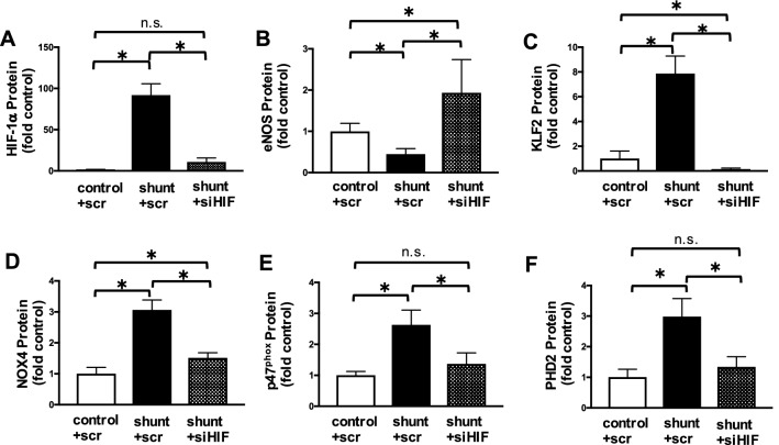
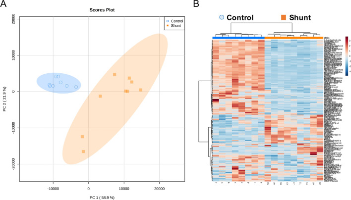
Normal growth and development of lymphatic structures depends on mechanical forces created by accumulating interstitial fluid. However, prolonged exposure to pathologic mechanical stimuli generated by chronically elevated lymph flow results in lymphatic dysfunction. The mechanisms that transduce these mechanical forces are not fully understood. Our objective was to investigate molecular mechanisms that alter the growth and metabolism of isolated lymphatic endothelial cells (LECs) exposed to prolonged pathologically elevated lymph flow in vivo within the anatomic and physiologic context of a large animal model of congenital heart disease with increased pulmonary blood flow using in vitro approaches. To this end, late gestation fetal lambs underwent in utero placement of an aortopulmonary graft (shunt). Four weeks after birth, LECs were isolated and cultured from control and shunt lambs. Redox status and proliferation were quantified, and transcriptional profiling and metabolomic analyses were performed. Shunt LECs exhibited hyperproliferative growth driven by increased levels of Hypoxia Inducible Factor 1α (HIF-1α), along with upregulated expression of known HIF-1α target genes in response to mechanical stimuli and shear stress. Compared to control LECs, shunt LECs exhibited abnormal metabolism including abnormalities of glycolysis, the TCA cycle and aerobic respiration. In conclusion, LECs from lambs exposed in vivo to chronically increased pulmonary lymph flow are hyperproliferative, have enhanced expression of HIF-1α and its target genes, and demonstrate altered central carbon metabolism in vitro. Importantly, these findings suggest provocative therapeutic targets for patients with lymphatic abnormalities.

摘要

正常的淋巴结构生长和发育依赖于由间质液积累产生的机械力。然而,长期暴露于由慢性升高的淋巴流量产生的病理机械刺激会导致淋巴功能障碍。将这些机械力转化为信号的机制尚未完全阐明。我们的目的是研究分子机制,即在先天性心脏病大动物模型中,在增加肺血流量的解剖和生理背景下,体内长时间暴露于病理升高的淋巴流量会改变分离的淋巴内皮细胞(LEC)的生长和代谢,使用体外方法。为此,在妊娠晚期,胎儿羔羊接受了宫内放置的主动脉肺动脉移植物(分流器)。出生后 4 周,从对照和分流羔羊中分离并培养 LEC。定量测定氧化还原状态和增殖,并进行转录谱和代谢组学分析。分流 LEC 表现出由缺氧诱导因子 1α(HIF-1α)水平升高驱动的过度增殖性生长,以及对机械刺激和切应力的已知 HIF-1α靶基因的上调表达。与对照 LEC 相比,分流 LEC 表现出异常代谢,包括糖酵解、三羧酸循环和有氧呼吸的异常。总之,体内暴露于慢性增加的肺淋巴流量的羔羊 LEC 过度增殖,HIF-1α及其靶基因表达增强,并在体外表现出中央碳代谢异常。重要的是,这些发现为淋巴异常患者提供了有前景的治疗靶点。

https://cdn.ncbi.nlm.nih.gov/pmc/blobs/5ba9/7809484/70169599a96c/41598_2020_80882_Fig7_HTML.jpg
https://cdn.ncbi.nlm.nih.gov/pmc/blobs/5ba9/7809484/4a37d5213e3d/41598_2020_80882_Fig1_HTML.jpg
https://cdn.ncbi.nlm.nih.gov/pmc/blobs/5ba9/7809484/dcd1832d8271/41598_2020_80882_Fig2_HTML.jpg
https://cdn.ncbi.nlm.nih.gov/pmc/blobs/5ba9/7809484/4bfd1882ec0c/41598_2020_80882_Fig3_HTML.jpg
https://cdn.ncbi.nlm.nih.gov/pmc/blobs/5ba9/7809484/125aee2492f0/41598_2020_80882_Fig4_HTML.jpg
https://cdn.ncbi.nlm.nih.gov/pmc/blobs/5ba9/7809484/162b4d4900e2/41598_2020_80882_Fig5_HTML.jpg
https://cdn.ncbi.nlm.nih.gov/pmc/blobs/5ba9/7809484/f68b6c7ee3ab/41598_2020_80882_Fig6_HTML.jpg
https://cdn.ncbi.nlm.nih.gov/pmc/blobs/5ba9/7809484/70169599a96c/41598_2020_80882_Fig7_HTML.jpg

相似文献

1
HIF-1α promotes cellular growth in lymphatic endothelial cells exposed to chronically elevated pulmonary lymph flow.低氧诱导因子 1α 促进慢性肺淋巴流量升高条件下淋巴管内皮细胞的增殖。
Sci Rep. 2021 Jan 14;11(1):1468. doi: 10.1038/s41598-020-80882-1.
2
KLF2-mediated disruption of PPAR-γ signaling in lymphatic endothelial cells exposed to chronically increased pulmonary lymph flow.KLF2 介导的慢性肺淋巴流量增加诱导的淋巴管内皮细胞中过氧化物酶体增殖物激活受体 γ 信号通路破坏。
Am J Physiol Heart Circ Physiol. 2018 Jul 1;315(1):H173-H181. doi: 10.1152/ajpheart.00635.2017. Epub 2018 Apr 6.
3
Disrupted NOS signaling in lymphatic endothelial cells exposed to chronically increased pulmonary lymph flow.慢性肺淋巴流量增加导致淋巴管内皮细胞中一氧化氮合酶信号转导紊乱。
Am J Physiol Heart Circ Physiol. 2016 Jul 1;311(1):H137-45. doi: 10.1152/ajpheart.00649.2015. Epub 2016 May 13.
4
Altered reactivity and nitric oxide signaling in the isolated thoracic duct from an ovine model of congenital heart disease with increased pulmonary blood flow.在一种肺动脉血流量增加的先天性心脏病绵羊模型的离体胸导管中,反应性和一氧化氮信号发生改变。
Am J Physiol Heart Circ Physiol. 2014 Apr 1;306(7):H954-62. doi: 10.1152/ajpheart.00841.2013. Epub 2014 Feb 14.
5
Altered hypoxia-inducible factor-1α (HIF-1α) signaling contributes to impaired angiogenesis in fetal lambs with persistent pulmonary hypertension of the newborn (PPHN).缺氧诱导因子-1α(HIF-1α)信号通路改变,导致患有新生儿持续性肺动脉高压(PPHN)的胎羊血管生成受损。
Physiol Rep. 2019 Feb;7(3):e13986. doi: 10.14814/phy2.13986.
6
Ovine Models of Congenital Heart Disease and the Consequences of Hemodynamic Alterations for Pulmonary Artery Remodeling.羊的先天性心脏病模型及血液动力改变对肺动脉重构的影响。
Am J Respir Cell Mol Biol. 2019 May;60(5):503-514. doi: 10.1165/rcmb.2018-0305MA.
7
Altered lymphatics in an ovine model of congenital heart disease with increased pulmonary blood flow.肺血流量增加的先天性心脏病羊模型中的淋巴管改变。
Am J Physiol Lung Cell Mol Physiol. 2012 Mar 15;302(6):L530-40. doi: 10.1152/ajplung.00324.2011. Epub 2011 Dec 29.
8
ORAI1 Activates Proliferation of Lymphatic Endothelial Cells in Response to Laminar Flow Through Krüppel-Like Factors 2 and 4.ORAI1通过Krüppel样因子2和4响应层流激活淋巴管内皮细胞增殖。
Circ Res. 2017 Apr 28;120(9):1426-1439. doi: 10.1161/CIRCRESAHA.116.309548. Epub 2017 Feb 6.
9
Hypoxia-inducible factors HIF-1alpha and HIF-2alpha are decreased in an experimental model of severe respiratory distress syndrome in preterm lambs.在早产羔羊严重呼吸窘迫综合征的实验模型中,缺氧诱导因子HIF-1α和HIF-2α减少。
Am J Physiol Lung Cell Mol Physiol. 2007 Jun;292(6):L1345-51. doi: 10.1152/ajplung.00372.2006. Epub 2007 Feb 16.
10
Breast cancer cells condition lymphatic endothelial cells within pre-metastatic niches to promote metastasis.乳腺癌细胞会影响前转移微环境中的淋巴管内皮细胞,以促进转移。
Nat Commun. 2014 Sep 2;5:4715. doi: 10.1038/ncomms5715.

引用本文的文献

1
Natural medicines target tumor vascular microenvironment to inhibit tumor.天然药物靶向肿瘤血管微环境以抑制肿瘤。
Genes Dis. 2025 Apr 8;12(6):101623. doi: 10.1016/j.gendis.2025.101623. eCollection 2025 Nov.
2
Mechanotransductive stabilization of HIF-1α is inhibited by mitochondrial antioxidant therapy in the setting of pulmonary overcirculation.在肺循环过度的情况下,线粒体抗氧化治疗可抑制HIF-1α的机械转导稳定性。
Sci Rep. 2025 May 10;15(1):16320. doi: 10.1038/s41598-025-99062-0.
3
Influence of Simulated Altitude Exposure (2500 m) on Patients with Fontan Palliation Based on Circulating Hypoxia-Associated Factors.

本文引用的文献

1
Mitochondria in the Pulmonary Vasculature in Health and Disease: Oxygen-Sensing, Metabolism, and Dynamics.肺部血管中的线粒体在健康和疾病中的作用:氧感应、代谢和动力学。
Compr Physiol. 2020 Mar 12;10(2):713-765. doi: 10.1002/cphy.c190027.
2
VE-Cadherin Is Required for Lymphatic Valve Formation and Maintenance.VE-钙黏蛋白对于淋巴管瓣膜的形成和维持是必需的。
Cell Rep. 2019 Aug 27;28(9):2397-2412.e4. doi: 10.1016/j.celrep.2019.07.072.
3
Mechanically activated ion channel PIEZO1 is required for lymphatic valve formation.机械激活的离子通道 PIEZO1 对于淋巴管瓣膜的形成是必需的。
基于循环缺氧相关因子的模拟海拔暴露(2500米)对接受Fontan姑息治疗患者的影响。
Int J Mol Sci. 2025 Jan 21;26(3):887. doi: 10.3390/ijms26030887.
4
MicroRNA in pediatric pulmonary hypertension microRNA profiling to inform disease classification, severity, and treatment response in pediatric pulmonary hypertension.小儿肺动脉高压中的微小RNA 微小RNA分析用于指导小儿肺动脉高压的疾病分类、严重程度及治疗反应
Am J Physiol Heart Circ Physiol. 2025 Jan 1;328(1):H47-H57. doi: 10.1152/ajpheart.00622.2024. Epub 2024 Nov 26.
5
Inflammatory Alterations to Renal Lymphatic Endothelial Cell Gene Expression in Mouse Models of Hypertension.高血压小鼠模型中肾脏淋巴内皮细胞基因表达的炎症改变。
Kidney Blood Press Res. 2024;49(1):588-604. doi: 10.1159/000539721. Epub 2024 Jul 22.
6
Lymphangiogenesis: A new strategy for heart disease treatment (Review).淋巴管生成:心脏病治疗的新策略(综述)。
Int J Mol Med. 2024 Apr;53(4). doi: 10.3892/ijmm.2024.5359. Epub 2024 Feb 23.
7
Molecular and metabolic orchestration of the lymphatic vasculature in physiology and pathology.淋巴血管系统在生理和病理中的分子和代谢调控。
Nat Commun. 2023 Dec 16;14(1):8389. doi: 10.1038/s41467-023-44133-x.
8
Respiratory mechanics and gas exchange in an ovine model of congenital heart disease with increased pulmonary blood flow and pressure.先天性心脏病绵羊模型中肺血流和压力增加时的呼吸力学与气体交换
Front Physiol. 2023 Jun 9;14:1188824. doi: 10.3389/fphys.2023.1188824. eCollection 2023.
9
Reactive Oxygen Species in Regulating Lymphangiogenesis and Lymphatic Function.活性氧在调节淋巴管生成和淋巴管功能中的作用。
Cells. 2022 May 26;11(11):1750. doi: 10.3390/cells11111750.
10
Transcription Factor Control of Lymphatic Quiescence and Maturation of Lymphatic Neovessels in Development and Physiology.转录因子对发育和生理过程中淋巴管静止及新生淋巴管成熟的调控
Front Physiol. 2021 Nov 2;12:672987. doi: 10.3389/fphys.2021.672987. eCollection 2021.
Proc Natl Acad Sci U S A. 2018 Dec 11;115(50):12817-12822. doi: 10.1073/pnas.1817070115. Epub 2018 Nov 27.
4
HIF-1α contributes to tube malformation of human lymphatic endothelial cells by upregulating VEGFR-3.缺氧诱导因子-1α通过上调血管内皮生长因子受体-3促进人淋巴管内皮细胞的管腔畸形。
Int J Oncol. 2019 Jan;54(1):139-151. doi: 10.3892/ijo.2018.4623. Epub 2018 Nov 5.
5
Regulatory mechanisms of hypoxia-inducible factor 1 activity: Two decades of knowledge.缺氧诱导因子1活性的调控机制:二十年的研究历程
Cancer Sci. 2018 Mar;109(3):560-571. doi: 10.1111/cas.13483. Epub 2018 Jan 27.
6
How glucose, glutamine and fatty acid metabolism shape blood and lymph vessel development.葡萄糖、谷氨酰胺和脂肪酸代谢如何塑造血管和淋巴管发育。
Dev Biol. 2019 Mar 1;447(1):90-102. doi: 10.1016/j.ydbio.2017.12.001. Epub 2017 Dec 7.
7
Mechanical Activation of Hypoxia-Inducible Factor 1α Drives Endothelial Dysfunction at Atheroprone Sites.缺氧诱导因子1α的机械激活驱动易患动脉粥样硬化部位的内皮功能障碍。
Arterioscler Thromb Vasc Biol. 2017 Nov;37(11):2087-2101. doi: 10.1161/ATVBAHA.117.309249. Epub 2017 Sep 7.
8
Flow pattern-dependent endothelial cell responses through transcriptional regulation.通过转录调控的流型依赖性内皮细胞反应。
Cell Cycle. 2017 Oct 18;16(20):1893-1901. doi: 10.1080/15384101.2017.1364324. Epub 2017 Aug 18.
9
Molecular Sensors of Blood Flow in Endothelial Cells.内皮细胞中血流的分子传感器。
Trends Mol Med. 2017 Sep;23(9):850-868. doi: 10.1016/j.molmed.2017.07.007. Epub 2017 Aug 12.
10
Maintenance of redox homeostasis by hypoxia-inducible factors.缺氧诱导因子对氧化还原稳态的维持
Redox Biol. 2017 Oct;13:331-335. doi: 10.1016/j.redox.2017.05.022. Epub 2017 May 31.