Kragh Kasper Nørskov, Alhede Maria, Kvich Lasse, Bjarnsholt Thomas
Costerton Biofilm Center, Department of Immunology and Microbiology, Faculty of Health Sciences University of Copenhagen, Blegdamsvej 3B, 2200, Copenhagen, Denmark.
Department of Clinical Microbiology, Henrik Harpestrengs Vej 4A, Rigshospitalet, 2100, Copenhagen, Denmark.
Biofilm. 2019 Sep 12;1:100006. doi: 10.1016/j.bioflm.2019.100006. eCollection 2019 Dec.
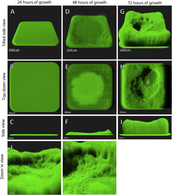
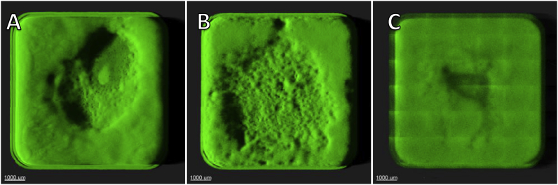
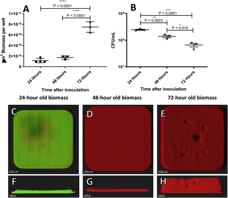
The microtiter assay is one of the most widely used methods for assessing biofilm formation. Though it has high throughput, this assay is known for its substantial deviation from experiment to experiment, and even from well to well. Since the assay constitutes one of the pillars of biofilm research, it was decided to examine the wells of a microtiter plate directly during growth, treatment, and the steps involved in crystal violet (CV) measurements. An inverted Zeiss LSM 880 confocal laser scanning microscope was used to visualize and quantify biomass directly in the wells of the microtiter plate. Green fluorescent protein-tagged PAO1, and live/dead stains were used to assess the structure, state, and position of biomass build-up. Microscopic observations were compared with colony-forming unit (CFU) and CV measurements. The development and the structured architecture of biomass was observed in real-time in the wells. Three-dimensional images of biomass were obtained from all of the microtiter wells; these showed variations from well to well. CV staining showed large variations in remaining biomass, depending on the method selected to remove the supernatant prior to CV staining (i.e. pipetting or manually discarding the fluid by inversion, washed or unwashed wells). Colony-forming unit counts or live/dead staining used to evaluate biomass with or without antibiotic treatment proved imprecise due to aggregation, limited removal of biomass, and overestimation of dead staining. The highly structured microenvironment of biomass in microtiter wells needs to be considered when designing and analyzing experiments. When using microtiter plates, stochastic variation due to growth and handling may lead to flawed conclusions. It is therefore recommended that this assay be used as a screening tool rather than as a stand-alone experimental tool.
微量滴定法是评估生物膜形成最广泛使用的方法之一。尽管它具有高通量,但该方法因实验间甚至孔间存在显著偏差而闻名。由于该方法是生物膜研究的支柱之一,因此决定在生长、处理以及结晶紫(CV)测量所涉及的步骤中直接检查微量滴定板的孔。使用倒置的蔡司LSM 880共聚焦激光扫描显微镜直接观察和量化微量滴定板孔中的生物量。使用绿色荧光蛋白标记的PAO1以及活/死染色剂来评估生物量积累的结构、状态和位置。将显微镜观察结果与菌落形成单位(CFU)和CV测量结果进行比较。实时观察了孔中生物量的发育和结构化结构。从所有微量滴定孔中获得了生物量的三维图像;这些图像显示孔与孔之间存在差异。CV染色显示,根据CV染色前选择去除上清液的方法(即移液或通过倒置手动丢弃液体、洗涤或未洗涤的孔),剩余生物量存在很大差异。由于聚集、生物量去除有限以及死染色的高估,用于评估有无抗生素处理的生物量的菌落形成单位计数或活/死染色被证明不准确。在设计和分析实验时,需要考虑微量滴定孔中生物量的高度结构化微环境。使用微量滴定板时,由于生长和操作导致的随机变化可能会得出有缺陷的结论。因此,建议将该方法用作筛选工具,而不是独立的实验工具。