• 文献检索
  • 文档翻译
  • 深度研究
  • 学术资讯
  • Suppr Zotero 插件Zotero 插件
  • 邀请有礼
  • 套餐&价格
  • 历史记录
应用&插件
Suppr Zotero 插件Zotero 插件浏览器插件Mac 客户端Windows 客户端微信小程序
定价
高级版会员购买积分包购买API积分包
服务
文献检索文档翻译深度研究API 文档MCP 服务
关于我们
关于 Suppr公司介绍联系我们用户协议隐私条款
关注我们

Suppr 超能文献

核心技术专利:CN118964589B侵权必究
粤ICP备2023148730 号-1Suppr @ 2026

文献检索

告别复杂PubMed语法,用中文像聊天一样搜索,搜遍4000万医学文献。AI智能推荐,让科研检索更轻松。

立即免费搜索

文件翻译

保留排版,准确专业,支持PDF/Word/PPT等文件格式,支持 12+语言互译。

免费翻译文档

深度研究

AI帮你快速写综述,25分钟生成高质量综述,智能提取关键信息,辅助科研写作。

立即免费体验

视网膜成像研究诊断 COVID-19:病例报告。

Retinal imaging study diagnoses in COVID-19: a case report.

机构信息

Department of Ophthalmology, Castilla La Mancha University, Albacete, Spain.

Puerta de Hierro-Majadahonda University Hospital, Madrid, Spain.

出版信息

J Med Case Rep. 2021 Jan 15;15(1):15. doi: 10.1186/s13256-020-02620-5.

DOI:10.1186/s13256-020-02620-5
PMID:33451352
原文链接:https://pmc.ncbi.nlm.nih.gov/articles/PMC7810099/
Abstract

BACKGROUND

Hyperreflective lesions at the level of ganglion cell (GCL) and inner plexiform retinal layers (IPL) by optical coherence tomography (OCT) and cotton wool spots in the examination of the eye fundus have recently been described as findings in patients with COVID-19 infection.

CASE REPORT

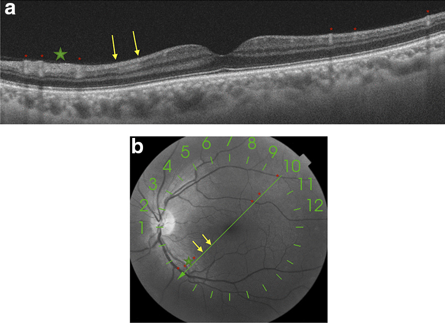
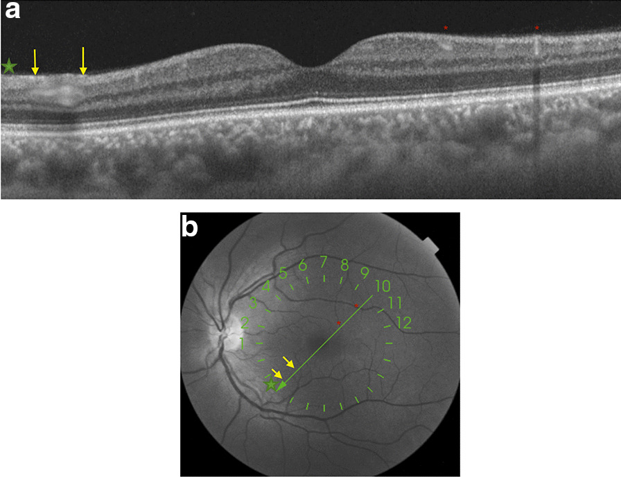
We report the case of a 42-year-old healthy Caucasian male anesthetist who had treated COVID-19 patients during the previous 5 weeks and suddenly presented with a temporal relative scotoma in his left eye. Best-corrected visual acuity was 20/20 for the left eye, and no discromatopsy or afferent pupillary defect was present. Visual field test was performed, with no significant findings associated with the focal loss of sensitivity described by the patient. The anterior segment was unremarkable on slit lamp examination in both eyes. Fundus examination of the left eye showed no significant findings. A placoid, hyperreflective band at the level of the GCL and IPL was visible in OCT which spared the outer retina, at the time of diagnosis and 1 month later. An oropharyngeal swab test was performed for severe acute respiratory syndrome coronavirus 2 (SARS-CoV-2) ribonucleic acid (RNA), immunoglobulin G (IgG) and immunoglobulin M (IgM) enzyme-linked immunosorbent assay (ELISA) determination. Real-time reverse-transcriptase polymerase chain reaction (RT-PCR) was negative. ELISA testing and a third rapid antibody detection test performed 7 days after the onset of symptoms were positive.

CONCLUSIONS

Ocular signs and symptoms in COVID-19 cases are rarely reported, but may be underestimated, especially those that affect the retina and occur in asymptomatic or paucisymptomatic cases. We present a case of COVID-19 diagnosis based on retinal ophthalmic examination.

摘要

背景

光学相干断层扫描(OCT)显示,新型冠状病毒肺炎(COVID-19)患者的神经节细胞(GCL)和内丛状视网膜层(IPL)水平出现高反射性病变,眼底检查可见棉絮斑。

病例报告

我们报告了 1 例 42 岁的健康白种男性麻醉师,他在过去 5 周内治疗了 COVID-19 患者,随后左眼出现颞侧相对暗点。左眼最佳矫正视力为 20/20,无辨色力障碍或传入性瞳孔缺陷。进行视野检查,与患者描述的局部敏感性丧失无关的显著发现。双眼裂隙灯检查前节未见明显异常。左眼眼底检查未见明显异常。OCT 显示,在诊断时和 1 个月后,左眼 GCL 和 IPL 水平可见盘状、高反射带,视网膜外层不受累。进行了严重急性呼吸综合征冠状病毒 2(SARS-CoV-2)核糖核酸(RNA)、免疫球蛋白 G(IgG)和免疫球蛋白 M(IgM)酶联免疫吸附试验(ELISA)测定的咽拭子检测。实时逆转录聚合酶链反应(RT-PCR)为阴性。ELISA 检测和发病后 7 天进行的第三次快速抗体检测均为阳性。

结论

COVID-19 病例中的眼部症状和体征很少报道,但可能被低估,尤其是那些影响视网膜且发生在无症状或症状轻微病例中的症状和体征。我们根据眼科检查诊断了 1 例 COVID-19 病例。

https://cdn.ncbi.nlm.nih.gov/pmc/blobs/1048/7811256/11fb182e29fe/13256_2020_2620_Fig2_HTML.jpg
https://cdn.ncbi.nlm.nih.gov/pmc/blobs/1048/7811256/933260b9bd1c/13256_2020_2620_Fig1_HTML.jpg
https://cdn.ncbi.nlm.nih.gov/pmc/blobs/1048/7811256/11fb182e29fe/13256_2020_2620_Fig2_HTML.jpg
https://cdn.ncbi.nlm.nih.gov/pmc/blobs/1048/7811256/933260b9bd1c/13256_2020_2620_Fig1_HTML.jpg
https://cdn.ncbi.nlm.nih.gov/pmc/blobs/1048/7811256/11fb182e29fe/13256_2020_2620_Fig2_HTML.jpg

相似文献

1
Retinal imaging study diagnoses in COVID-19: a case report.视网膜成像研究诊断 COVID-19:病例报告。
J Med Case Rep. 2021 Jan 15;15(1):15. doi: 10.1186/s13256-020-02620-5.
2
Retinal microvascular abnormalities, cotton wool-like lesions, and macular edema following COVID-19 in a patient previously vaccinated with AstraZeneca and idiopathic myopathy.患者此前接种过阿斯利康疫苗,患有特发性肌病,感染 COVID-19 后出现视网膜微血管异常、棉絮状斑和黄斑水肿。
Eur J Ophthalmol. 2023 Sep;33(5):NP55-NP59. doi: 10.1177/11206721221130393. Epub 2022 Oct 13.
3
Covid-19-Associated Retinopathy: A Case Report.Covid-19 相关视网膜病变:病例报告。
Ocul Immunol Inflamm. 2020 Nov 16;28(8):1293-1297. doi: 10.1080/09273948.2020.1825751. Epub 2020 Oct 6.
4
Symptomatic Retinal Microangiopathy in a Patient with Coronavirus Disease 2019 (COVID-19): Single Case Report.COVID-19 相关的有症状视网膜微血管病变:单个病例报告。
Ocul Immunol Inflamm. 2021 May 19;29(4):642-644. doi: 10.1080/09273948.2020.1852260. Epub 2020 Dec 7.
5
Vitritis and Outer Retinal Abnormalities in a Patient with COVID-19.COVID-19 患者的虹膜炎和外视网膜异常。
Ocul Immunol Inflamm. 2020 Nov 16;28(8):1298-1300. doi: 10.1080/09273948.2020.1821898. Epub 2020 Oct 6.
6
Acute Zonal Cone Photoreceptor Outer Segment Loss.急性区域性锥体细胞光感受器外段缺失
JAMA Ophthalmol. 2017 May 1;135(5):487-490. doi: 10.1001/jamaophthalmol.2017.0451.
7
The characteristics of acute macular neuroretinopathy following COVID-19 infection.急性黄斑神经视网膜病变的特点,继 COVID-19 感染之后。
BMC Ophthalmol. 2024 Jan 10;24(1):19. doi: 10.1186/s12886-024-03283-2.
8
CLINICALLY INVISIBLE RETINAL HEMANGIOBLASTOMAS DETECTED BY SPECTRAL DOMAIN OPTICAL COHERENCE TOMOGRAPHY AND FLUORESCEIN ANGIOGRAPHY IN TWINS.通过光谱域光学相干断层扫描和荧光素血管造影在双胞胎中检测到的临床隐匿性视网膜血管母细胞瘤
Retin Cases Brief Rep. 2018;12(1):12-16. doi: 10.1097/ICB.0000000000000382.
9
A case of retinal vascular involvement in a 6-year-old patient with Covid-19.一名6岁新冠病毒肺炎患者出现视网膜血管受累的病例。
Eur J Ophthalmol. 2022 Jul;32(4):NP1-NP5. doi: 10.1177/11206721211027069. Epub 2021 Jun 26.
10
Overview of the retina and imaging in patients with severe acute respiratory syndrome coronavirus 2.严重急性呼吸综合征冠状病毒 2 患者的视网膜和成像概述。
Int Ophthalmol. 2022 Nov;42(11):3601-3610. doi: 10.1007/s10792-022-02338-x. Epub 2022 May 11.

引用本文的文献

1
Manifestations of COVID-19 in the posterior eye segment - Up-to-date.新型冠状病毒肺炎在后段眼的表现——最新情况。
Oman J Ophthalmol. 2024 Jun 27;17(2):166-172. doi: 10.4103/ojo.ojo_212_22. eCollection 2024 May-Aug.
2
Outer Retinopathies Associated with COVID-19 Infection: Case Reports and Review of Literature.与新型冠状病毒肺炎感染相关的外层视网膜病变:病例报告及文献综述
Case Rep Ophthalmol Med. 2024 Mar 8;2024:7227086. doi: 10.1155/2024/7227086. eCollection 2024.
3
Ophthalmic Presentations and Manifestations of COVID-19: A Systematic Review of Global Observations.

本文引用的文献

1
Neuro-ophthalmologic complications of coronavirus disease 2019 (COVID-19).新型冠状病毒肺炎(COVID-19)的神经眼科并发症。
Neurosci Lett. 2021 Jan 18;742:135531. doi: 10.1016/j.neulet.2020.135531. Epub 2020 Nov 25.
2
Human coronaviruses: ophthalmic manifestations.人类冠状病毒:眼部表现
BMJ Open Ophthalmol. 2020 Nov 2;5(1):e000630. doi: 10.1136/bmjophth-2020-000630. eCollection 2020.
3
COVID-19 retinal microangiopathy as an in vivo biomarker of systemic vascular disease?COVID-19 视网膜微血管病变是否可作为系统性血管疾病的体内生物标志物?
2019冠状病毒病的眼科表现与临床表现:全球观察的系统评价
Cureus. 2023 Jun 20;15(6):e40695. doi: 10.7759/cureus.40695. eCollection 2023 Jun.
4
Update on overview of ocular manifestations of COVID-19.新型冠状病毒肺炎眼部表现概述的最新进展。
Front Med (Lausanne). 2022 Sep 13;9:877023. doi: 10.3389/fmed.2022.877023. eCollection 2022.
5
Use of Imaging Technology to Assess the Effect of COVID-19 on Retinal Tissues: A Systematic Review.使用成像技术评估2019冠状病毒病对视网膜组织的影响:一项系统评价
Ophthalmol Ther. 2022 Jun;11(3):1017-1030. doi: 10.1007/s40123-022-00509-8. Epub 2022 Apr 29.
6
Angiotensin Converting Enzyme 2 (ACE2) Expression in the Aged Brain and Visual System.血管紧张素转换酶2(ACE2)在老年大脑和视觉系统中的表达。
J Aging Sci. 2021;Vol 9(Suppl 7). Epub 2021 Sep 30.
7
Vogt-Koyanagi-Harada Disease Following COVID-19 Infection.新型冠状病毒肺炎感染后发生的伏格特-小柳-原田病
Case Rep Ophthalmol. 2021 Sep 21;12(3):804-808. doi: 10.1159/000518834. eCollection 2021 Sep-Dec.
8
Vitreoretinal abnormalities in corona virus disease 2019 patients: What we know so far.2019冠状病毒病患者的玻璃体视网膜异常:我们目前所了解的情况。
Taiwan J Ophthalmol. 2021 Aug 28;11(3):232-243. doi: 10.4103/tjo.tjo_30_21. eCollection 2021 Jul-Sep.
9
COVID-19-Related Retinal Micro-vasculopathy - A Review of Current Evidence.COVID-19 相关视网膜微血管病变——现有证据的综述。
Am J Ophthalmol. 2022 Mar;235:98-110. doi: 10.1016/j.ajo.2021.09.019. Epub 2021 Sep 26.
J Intern Med. 2021 Jan;289(1):116-120. doi: 10.1111/joim.13156. Epub 2020 Jul 30.
4
Concerns about the interpretation of OCT and fundus findings in COVID-19 patients in recent Lancet publication.近期《柳叶刀》发表的文章对新冠病毒肺炎患者光学相干断层扫描(OCT)及眼底检查结果的解读提出了担忧。
Eye (Lond). 2020 Dec;34(12):2153-2154. doi: 10.1038/s41433-020-1084-9. Epub 2020 Jul 9.
5
Cerebrovascular disease in patients with COVID-19: neuroimaging, histological and clinical description.COVID-19 患者的脑血管病:神经影像学、组织学和临床描述。
Brain. 2020 Oct 1;143(10):3089-3103. doi: 10.1093/brain/awaa239.
6
Retinal findings in patients with COVID-19.新型冠状病毒肺炎患者的视网膜病变
Lancet. 2020 May 23;395(10237):1610. doi: 10.1016/S0140-6736(20)31014-X. Epub 2020 May 12.
7
Hyperinflammation and derangement of renin-angiotensin-aldosterone system in COVID-19: A novel hypothesis for clinically suspected hypercoagulopathy and microvascular immunothrombosis.新型冠状病毒肺炎中过度炎症反应和肾素-血管紧张素-醛固酮系统紊乱:疑似高凝状态和微血管免疫血栓形成的新假说。
Clin Chim Acta. 2020 Aug;507:167-173. doi: 10.1016/j.cca.2020.04.027. Epub 2020 Apr 26.
8
A first case of meningitis/encephalitis associated with SARS-Coronavirus-2.首例与 SARS-CoV-2 相关的脑膜炎/脑炎。
Int J Infect Dis. 2020 May;94:55-58. doi: 10.1016/j.ijid.2020.03.062. Epub 2020 Apr 3.
9
Characteristics of Ocular Findings of Patients With Coronavirus Disease 2019 (COVID-19) in Hubei Province, China.中国湖北省 2019 年冠状病毒病(COVID-19)患者的眼部表现特征。
JAMA Ophthalmol. 2020 May 1;138(5):575-578. doi: 10.1001/jamaophthalmol.2020.1291.
10
Spectrum of Retinal Vascular Diseases Associated With Paracentral Acute Middle Maculopathy.与中心旁急性黄斑病变相关的视网膜血管疾病谱
Am J Ophthalmol. 2015 Jul;160(1):26-34.e1. doi: 10.1016/j.ajo.2015.04.004. Epub 2015 Apr 4.