• 文献检索
  • 文档翻译
  • 深度研究
  • 学术资讯
  • Suppr Zotero 插件Zotero 插件
  • 邀请有礼
  • 套餐&价格
  • 历史记录
应用&插件
Suppr Zotero 插件Zotero 插件浏览器插件Mac 客户端Windows 客户端微信小程序
定价
高级版会员购买积分包购买API积分包
服务
文献检索文档翻译深度研究API 文档MCP 服务
关于我们
关于 Suppr公司介绍联系我们用户协议隐私条款
关注我们

Suppr 超能文献

核心技术专利:CN118964589B侵权必究
粤ICP备2023148730 号-1Suppr @ 2026

文献检索

告别复杂PubMed语法,用中文像聊天一样搜索,搜遍4000万医学文献。AI智能推荐,让科研检索更轻松。

立即免费搜索

文件翻译

保留排版,准确专业,支持PDF/Word/PPT等文件格式,支持 12+语言互译。

免费翻译文档

深度研究

AI帮你快速写综述,25分钟生成高质量综述,智能提取关键信息,辅助科研写作。

立即免费体验

特征歧义缓解算子模型有助于提高 X 射线照片中的骨折检测。

The Feature Ambiguity Mitigate Operator model helps improve bone fracture detection on X-ray radiograph.

机构信息

Department of Radiology, Third Hospital of Hebei Medical University, Hebei Province, Shijiazhuang, 050000, People's Republic of China.

DeepWise AI Lab, Beijing, People's Republic of China.

出版信息

Sci Rep. 2021 Jan 15;11(1):1589. doi: 10.1038/s41598-021-81236-1.

DOI:10.1038/s41598-021-81236-1
PMID:33452403
原文链接:https://pmc.ncbi.nlm.nih.gov/articles/PMC7810849/
Abstract

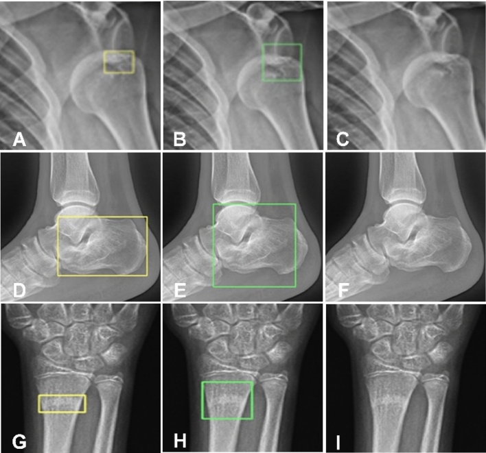
This study was performed to propose a method, the Feature Ambiguity Mitigate Operator (FAMO) model, to mitigate feature ambiguity in bone fracture detection on radiographs of various body parts. A total of 9040 radiographic studies were extracted. These images were classified into several body part types including 1651 hand, 1302 wrist, 406 elbow, 696 shoulder, 1580 pelvic, 948 knee, 1180 ankle, and 1277 foot images. Instance segmentation was annotated by radiologists. The ResNext-101+FPN was employed as the baseline network structure and the FAMO model for processing. The proposed FAMO model and other ablative models were tested on a test set of 20% total radiographs in a balanced body part distribution. To the per-fracture extent, an AP (average precision) analysis was performed. For per-image and per-case, the sensitivity, specificity, and AUC (area under the receiver operating characteristic curve) were analyzed. At the per-fracture level, the controlled experiment set the baseline AP to 76.8% (95% CI: 76.1%, 77.4%), and the major experiment using FAMO as a preprocessor improved the AP to 77.4% (95% CI: 76.6%, 78.2%). At the per-image level, the sensitivity, specificity, and AUC were 61.9% (95% CI: 58.7%, 65.0%), 91.5% (95% CI: 89.5%, 93.3%), and 74.9% (95% CI: 74.1%, 75.7%), respectively, for the controlled experiment, and 64.5% (95% CI: 61.3%, 67.5%), 92.9% (95% CI: 91.0%, 94.5%), and 77.5% (95% CI: 76.5%, 78.5%), respectively, for the experiment with FAMO. At the per-case level, the sensitivity, specificity, and AUC were 74.9% (95% CI: 70.6%, 78.7%), 91.7%% (95% CI: 88.8%, 93.9%), and 85.7% (95% CI: 84.8%, 86.5%), respectively, for the controlled experiment, and 77.5% (95% CI: 73.3%, 81.1%), 93.4% (95% CI: 90.7%, 95.4%), and 86.5% (95% CI: 85.6%, 87.4%), respectively, for the experiment with FAMO. In conclusion, in bone fracture detection, FAMO is an effective preprocessor to enhance model performance by mitigating feature ambiguity in the network.

摘要

本研究旨在提出一种方法,即特征歧义缓解算子(FAMO)模型,以减轻各种身体部位 X 光片上骨折检测中的特征歧义。共提取了 9040 项放射学研究。这些图像被分为几种身体部位类型,包括 1651 只手、1302 只手腕、406 只肘部、696 只肩部、1580 只骨盆、948 只膝盖、1180 只脚踝和 1277 只脚图像。由放射科医生对实例分割进行注释。采用 ResNext-101+FPN 作为基线网络结构,采用 FAMO 模型进行处理。在平衡身体部位分布的 20%总 X 光片的测试集中测试了所提出的 FAMO 模型和其他消融模型。对于每个骨折程度,进行了平均精度(AP)分析。对于每张图像和每个病例,分析了敏感性、特异性和 AUC(接收器工作特征曲线下的面积)。在骨折程度上,对照实验将基线 AP 设置为 76.8%(95%CI:76.1%,77.4%),而主要实验使用 FAMO 作为预处理器将 AP 提高到 77.4%(95%CI:76.6%,78.2%)。在图像级别,对照实验的敏感性、特异性和 AUC 分别为 61.9%(95%CI:58.7%,65.0%)、91.5%(95%CI:89.5%,93.3%)和 74.9%(95%CI:74.1%,75.7%),而 FAMO 实验的敏感性、特异性和 AUC 分别为 64.5%(95%CI:61.3%,67.5%)、92.9%(95%CI:91.0%,94.5%)和 77.5%(95%CI:76.5%,78.5%)。在病例级别,对照实验的敏感性、特异性和 AUC 分别为 74.9%(95%CI:70.6%,78.7%)、91.7%(95%CI:88.8%,93.9%)和 85.7%(95%CI:84.8%,86.5%),而 FAMO 实验的敏感性、特异性和 AUC 分别为 77.5%(95%CI:73.3%,81.1%)、93.4%(95%CI:90.7%,95.4%)和 86.5%(95%CI:85.6%,87.4%)。总之,在骨折检测中,FAMO 是一种有效的预处理方法,可以通过减轻网络中的特征歧义来提高模型性能。

https://cdn.ncbi.nlm.nih.gov/pmc/blobs/b144/7810849/cf01545b550a/41598_2021_81236_Fig7_HTML.jpg
https://cdn.ncbi.nlm.nih.gov/pmc/blobs/b144/7810849/9d45e370d225/41598_2021_81236_Fig1_HTML.jpg
https://cdn.ncbi.nlm.nih.gov/pmc/blobs/b144/7810849/367ded8dd215/41598_2021_81236_Fig2_HTML.jpg
https://cdn.ncbi.nlm.nih.gov/pmc/blobs/b144/7810849/40cf6f1e1ef3/41598_2021_81236_Fig3_HTML.jpg
https://cdn.ncbi.nlm.nih.gov/pmc/blobs/b144/7810849/b9acea039b4c/41598_2021_81236_Fig4_HTML.jpg
https://cdn.ncbi.nlm.nih.gov/pmc/blobs/b144/7810849/69bf316ca24e/41598_2021_81236_Fig5_HTML.jpg
https://cdn.ncbi.nlm.nih.gov/pmc/blobs/b144/7810849/3568d9d9f54e/41598_2021_81236_Fig6_HTML.jpg
https://cdn.ncbi.nlm.nih.gov/pmc/blobs/b144/7810849/cf01545b550a/41598_2021_81236_Fig7_HTML.jpg

相似文献

1
The Feature Ambiguity Mitigate Operator model helps improve bone fracture detection on X-ray radiograph.特征歧义缓解算子模型有助于提高 X 射线照片中的骨折检测。
Sci Rep. 2021 Jan 15;11(1):1589. doi: 10.1038/s41598-021-81236-1.
2
Using a Dual-Input Convolutional Neural Network for Automated Detection of Pediatric Supracondylar Fracture on Conventional Radiography.利用双输入卷积神经网络自动检测常规 X 光片中的小儿髁上骨折。
Invest Radiol. 2020 Feb;55(2):101-110. doi: 10.1097/RLI.0000000000000615.
3
Deep Learning Assistance Closes the Accuracy Gap in Fracture Detection Across Clinician Types.深度学习辅助缩小了不同临床医生类型在骨折检测中的准确性差距。
Clin Orthop Relat Res. 2023 Mar 1;481(3):580-588. doi: 10.1097/CORR.0000000000002385. Epub 2022 Sep 9.
4
Improving Radiographic Fracture Recognition Performance and Efficiency Using Artificial Intelligence.利用人工智能提高放射科骨折识别性能和效率。
Radiology. 2022 Mar;302(3):627-636. doi: 10.1148/radiol.210937. Epub 2021 Dec 21.
5
Deep learning evaluation of pelvic radiographs for position, hardware presence, and fracture detection.深度学习评估骨盆 X 光片的位置、硬件存在情况和骨折检测。
Eur J Radiol. 2020 Sep;130:109139. doi: 10.1016/j.ejrad.2020.109139. Epub 2020 Jun 21.
6
Comparison of Chest Radiograph Interpretations by Artificial Intelligence Algorithm vs Radiology Residents.人工智能算法与放射科住院医师对胸部 X 线片解读的比较。
JAMA Netw Open. 2020 Oct 1;3(10):e2022779. doi: 10.1001/jamanetworkopen.2020.22779.
7
Automated detection of moderate and large pneumothorax on frontal chest X-rays using deep convolutional neural networks: A retrospective study.使用深度卷积神经网络自动检测正位胸部 X 光片中的中至大量气胸:一项回顾性研究。
PLoS Med. 2018 Nov 20;15(11):e1002697. doi: 10.1371/journal.pmed.1002697. eCollection 2018 Nov.
8
Convolutional Neural Networks for Automated Fracture Detection and Localization on Wrist Radiographs.用于腕部X光片骨折自动检测与定位的卷积神经网络
Radiol Artif Intell. 2019 Jan 30;1(1):e180001. doi: 10.1148/ryai.2019180001. eCollection 2019 Jan.
9
Development and Validation of a Convolutional Neural Network Model to Predict a Pathologic Fracture in the Proximal Femur Using Abdomen and Pelvis CT Images of Patients With Advanced Cancer.利用晚期癌症患者腹部和骨盆 CT 图像建立卷积神经网络模型预测股骨近端病理性骨折的研究
Clin Orthop Relat Res. 2023 Nov 1;481(11):2247-2256. doi: 10.1097/CORR.0000000000002771. Epub 2023 Aug 23.
10
Artificial intelligence to identify fractures on pediatric and young adult upper extremity radiographs.人工智能识别小儿和青年上肢 X 光片骨折。
Pediatr Radiol. 2023 Nov;53(12):2386-2397. doi: 10.1007/s00247-023-05754-y. Epub 2023 Sep 23.

引用本文的文献

1
Interpretable Severity Scoring of Pelvic Trauma Through Automated Fracture Detection and Bayesian Inference.通过自动骨折检测和贝叶斯推理对骨盆创伤进行可解释的严重程度评分。
IEEE Trans Med Imaging. 2025 Jan;44(1):130-141. doi: 10.1109/TMI.2024.3428836. Epub 2025 Jan 2.
2
Advances in Deep Learning-Based Medical Image Analysis.基于深度学习的医学图像分析进展
Health Data Sci. 2021 May 19;2021:8786793. doi: 10.34133/2021/8786793. eCollection 2021.
3
Fracture detection in pediatric wrist trauma X-ray images using YOLOv8 algorithm.

本文引用的文献

1
Convolutional Neural Networks for Automated Fracture Detection and Localization on Wrist Radiographs.用于腕部X光片骨折自动检测与定位的卷积神经网络
Radiol Artif Intell. 2019 Jan 30;1(1):e180001. doi: 10.1148/ryai.2019180001. eCollection 2019 Jan.
2
Ankle Fracture Detection Utilizing a Convolutional Neural Network Ensemble Implemented with a Small Sample, De Novo Training, and Multiview Incorporation.利用卷积神经网络集成进行小样本、从头训练和多视图合并实现的踝关节骨折检测。
J Digit Imaging. 2019 Aug;32(4):672-677. doi: 10.1007/s10278-018-0167-7.
3
Deep neural network improves fracture detection by clinicians.
使用 YOLOv8 算法检测小儿腕部创伤 X 射线图像中的骨折。
Sci Rep. 2023 Nov 16;13(1):20077. doi: 10.1038/s41598-023-47460-7.
4
A Pyramid Deep Feature Extraction Model for the Automatic Classification of Upper Extremity Fractures.一种用于上肢骨折自动分类的金字塔深度特征提取模型。
Diagnostics (Basel). 2023 Oct 26;13(21):3317. doi: 10.3390/diagnostics13213317.
5
Skeletal Fracture Detection with Deep Learning: A Comprehensive Review.基于深度学习的骨骼骨折检测:全面综述
Diagnostics (Basel). 2023 Oct 18;13(20):3245. doi: 10.3390/diagnostics13203245.
6
Artificial intelligence versus radiologist in the accuracy of fracture detection based on computed tomography images: a multi-dimensional, multi-region analysis.基于计算机断层扫描图像的骨折检测准确性:人工智能与放射科医生的多维、多区域分析
Quant Imaging Med Surg. 2023 Oct 1;13(10):6424-6433. doi: 10.21037/qims-23-428. Epub 2023 Sep 4.
7
Preoperative computed tomography morphological characteristics of displaced femoral neck fractures.术前 CT 形态学特征对移位型股骨颈骨折的诊断价值
Jt Dis Relat Surg. 2023 May 12;34(2):253-264. doi: 10.52312/jdrs.2023.1003.
8
Fracture Detection in Wrist X-ray Images Using Deep Learning-Based Object Detection Models.基于深度学习的目标检测模型在腕部 X 射线图像中的骨折检测。
Sensors (Basel). 2022 Feb 8;22(3):1285. doi: 10.3390/s22031285.
深度学习神经网络可帮助临床医生提高骨折检出率。
Proc Natl Acad Sci U S A. 2018 Nov 6;115(45):11591-11596. doi: 10.1073/pnas.1806905115. Epub 2018 Oct 22.
4
Artificial intelligence for analyzing orthopedic trauma radiographs.用于分析骨科创伤X光片的人工智能
Acta Orthop. 2017 Dec;88(6):581-586. doi: 10.1080/17453674.2017.1344459. Epub 2017 Jul 6.